Download

1 / 15
150 likes | 283 Views
Detection of Amnionless Protein in Kidney Sections. Authors Natasha Tonge Dr. Roberta Rivi. Acknowledgements. Dr. Roberta Rivi Dr. Sat Bhattacharya Dr. Katia Manova Dr. Betul Altay-Ozer Harlem Children Society Elizabeth Lacy Lab MSKCC. Wax in a Different Context.

E N D
Detection of Amnionless Protein in Kidney Sections Authors Natasha Tonge Dr. Roberta Rivi
Acknowledgements Dr. Roberta Rivi Dr. Sat Bhattacharya Dr. Katia Manova Dr. Betul Altay-Ozer Harlem Children Society Elizabeth Lacy Lab MSKCC
Wax in a Different Context Tissue sample is embedded in paraffin wax
Wax in a Different Context Tissue sample embedded in paraffin wax Sliced with microtome
Wax in a Different Context Tissue sample embedded in paraffin wax Sliced with microtome Placed in Tissue bath
Wax in a Different Context Tissue sample embedded in paraffin wax Sliced with microtome Placed in Tissue bath Mounted on a slide
Wax in a Different Context Tissue sample embedded in paraffin wax Sliced with microtome Placed in Tissue bath Mounted on a slide AutomaticallyProcessed
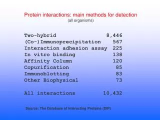
Antibodies in a Different Context Immunohistochemistry - a process used to detect proteins in a tissue section. Antigen - a substance that provokes an immune response Antibody – the protein complex that binds to antigens Enzyme - A protein that speeds up a chemical reaction. Substrate - The chemical that induces the chemical reaction
Antibodies in a Different Context Immunohistochemistry - a process used to detect proteins in a tissue section. Antigen - a substance that provokes an immune response Antibody – the protein complex that binds to antigens Enzyme - A protein that speeds up a chemical reaction. Substrate - The chemical that induces the chemical reaction primary antibody secondary antibody enzyme substrate Antigen
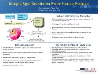
The Anatomy of the Kidney Ammnionless - protein located on the proximal tubule. Proximal Tubule - close to the glomerulus. Cortex - outer region of the kidney. Most likely to find the glomerulus and proximal tubule here. Medula - inner region of the kidney. Lacks ammnionless.
The Past and Future of this Experiment To Detect Amnionless in Kidney Sections: • Tissue samples are sectioned, placed on slides and stained • The staining process, immunohistochemistry, makes use of antibodies to induce a color change • That color change enabled us to see the location of the proximal tubules in the kidney sections. In future experiments the same process will be used to pinpoint mutations in abnormal mouse tissue.
Bibliography http://upload.wikimedia.org/wikipedia/en/thumb/b/b0/Kidney_section.jpg/180px-Kidney_section.jpg (altered by Natasha Tonge) http://mskweb3.mskcc.org/MolecularCytology/mccf-final.htm http://en.wikipedia.org/wiki/Human_hair