Download
1 / 91
920 likes | 1.19k Views
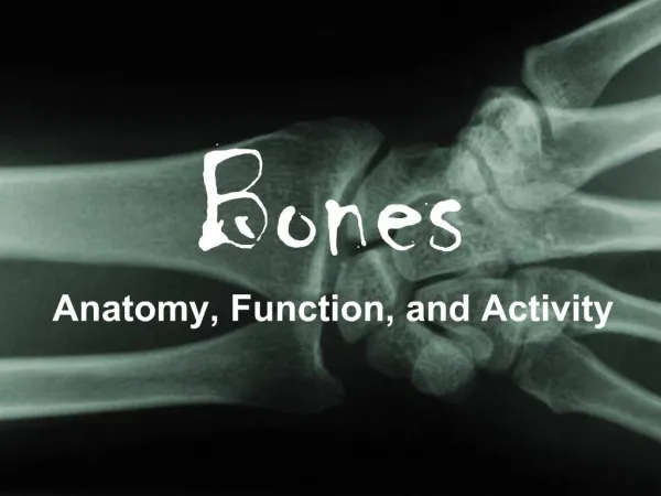
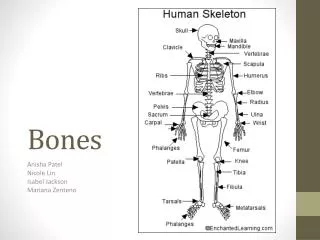
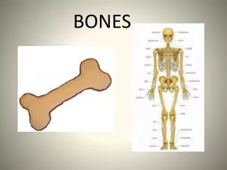
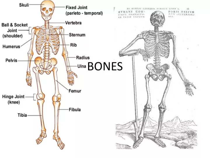
BONES. Osteology = study of bones. Tissue: Connective Functions: Support Protect soft tissue Points of attachment for muscles House blood producing cells Store inorganic salts. 206 bones 2 main divisions Axial Appendicular. Axial. Head, Neck, Trunk Skull Hyoid bone

E N D
Tissue: Connective • Functions: • Support • Protect soft tissue • Points of attachment for muscles • House blood producing cells • Store inorganic salts
206 bones • 2 main divisions • Axial • Appendicular
Axial • Head, Neck, Trunk • Skull • Hyoid bone • Vertebral column • Thoracic Cage (ribs, 12 pairs) • Sternum
APPENDICULAR • limbs and bones connecting the limbs to the: PECTORAL GIRDLE (scapula & clavicle), UPPER LIMBS (arms)PELVIC GIRDLE (coxal bones), LOWER LIMBS (legs)
Bone Classification • Long bones • Short bones • Flat bones • Irregular bones * Sesamoid bones
1. Long Bones- have long longitudinal axes ad expanded ends • EX: forearm, thigh bones 2. Short Bones- cube like with lengths/ widths roughly equal • Ex: wrist, ankles
3. Flat Bones- platelike structures with broad surfaces Ex: Ribs, scapulae
4. Irregular bones- variety of shapes, connected to several bones Ex: vertebrae, facial bones 5. Sesamoid or round bones- small and nodular Ex: kneecap
Long Bone • Epiphysis- expanded portion at each end of bone, articulates (forms a joint) • Diaphysis- shaft of bone • Articular cartilage- layer of hyaline cartilage • Proximal epiphysis – nearest to torso • Distal epiphysis- end furthest from torso • Periosteum- bone covered in a tough vascular covering of fibrous tissue
pg 194
Processes- bony projections for sites of attachment • Provide attachment sites, grooves/openings for passageways of blood vessels and nerves
2 Bone Types • Compact bone (Cortical) - tightly packed tissue, solid, strong - Wall of diaphysis • Spongy Bone (cancellous) - many branching bone plates, covered with a layer of compact bone.
Medullar Cavity- hollow chamber within the diaphysis connects to spaces in spongy bone. Filled with soft specialized tissue called bone marrow. • Endosteum- thin membrane containing bone-forming cells lining medullar cavity
Marrow- Red Marrow - mainly in spongy bone in adults. Produces blood cells Yellow Marrow - fat storage. Replaces much of the red marrow in diaphysis through childhood
Microscopic Structures pg 196 • Bone Extracellular Matrix = collagen / inorganic salts • Osteocytes- located in tiny bony chambers called lacunae • Transport nutrients and waste
Compact Bone • Osteon- (harversian system) cylinder unit around central canal • Contain blood vessels, nerve tissue, loose CT • Central canals – extend longitudinally through bone • Perforating canal (Volkmann’s canals)- transverse canals connect central canals • Contain blood vessels and nerves
Spongy bone • Cells lie within trabeculae • Nutrients from subs. diffusing into the canaliculi
Two types of bones based on development • 1. INTRAMEMBRANOUS BONES = broad, flat bones of the skull. • form from membrane-like sheets of connective tissue • 2. ENDOCHONDRAL BONES =masses of cartilage that are later replaced by bone tissue • EX: long bones
Intramembranous Bones • Osteoblasts appear in CT • Bone forming cells • Fibers appear in matrix • Calcification occurs • Deposits of salts in matrix • Osteoblasts become osteocytes
Endochondral • Skeleton in cartilage • Bone replaces cartilage • Ossification centers • Areas where bone formation starts • Blood vessels penetrate cartilage
ALL BONES START AS HYALINE CARTILAGE, areas gradually turn to bone • PRIMARY OSSIFICATION CENTER (shaft) • SECONDARY OSSIFICATION CENTER (ends)
Epiphyseal disk (growth plate) is a band of cartilage b/w the epiphysis and diaphysis • These areas increase bone length as the cells ossify • Cartilage becomes osteoblasts become osteocytes
RESORPTION • OSTEOCLASTS - dissolve bone tissue to release minerals, process is called RESORPTION
Factors Affecting Bone Growth • Absence of Calcium • Deforms bones • children= rickets • Adults= osteomalacia
Lack/Excess of growth hormone • Lack - Child= pituitary dwarfism • Excess- child= pituitary gigantism
Stress- causes bones to grow, lack of exercise causes bone tissue to waste away
Axial • Skull Cranium and facial bones • Hyoid bone- floats, helps with swallowing/ supports tongue • Vertebral Column • Sacrum • Coccyx • Thoracic Cage- 12 pairs of ribs and sternum
Appendicular • Pectoral girdle- scapula • Clavicle • Upper limbs- humerous, radius, ulna, carpals, metacarpals, phalanges • Pelvic girdle- coxa, pelvis • Lower Limbs- femur, tibia(large), fibula(slender), patella, tarsals, metatarsals, phalanges
3 Basic Types of Joints (articulations): 1. SYNARTHROTIC – immoveable joint, such as bones in the skull, these junctions are called SUTURES 2. AMPHIARTHOTIC – slightly moveable joint, vertebrae 3. DIARTHROTIC – freely moveable joint, such as shoulders, hips, knees, elbows, wrists, fingers… --these joints are enclosed within a fibrous capsule which contains a lubricating fluid called SYNOVIAL fluid. These are called SYNOVIAL JOINTS.
Cranium Bones 1. Frontal - anterior portion above eyes 2. Parietal – one on each side of the skull, just behind frontal bone 3. Occipital – forms the back of the skull and base of the cranium 4. Temporal – forms parts of the sides and base of cranium • Sphenoid – wedged between several other bones in anterior portion of the cranium • Ethomoid – form roof of nasal cavity
Sutures • 1. Coronal – between frontal and parietal bones • 2. Lambdoidal – between occipital and parietal bones • 3. Squamous – between temporal and parietal bones • 4. Sagittal - between parietal bones
Facial Bones immovable and 1 movable jawbone • Maxillary bones • Palatine bones • Zygomatic bones • Lacrimal bones • Nasal bones • Vomer bones • Inferior nasal conchae • mandible
Foremens • Allow blood vessels/nerves to travel through bone • Supraorbital foremen • Infraorbital foremen • Mental foremen