Download
1 / 73
740 likes | 961 Views
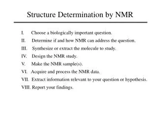
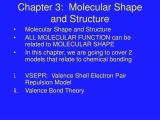
3 - NMR Data and Molecular Structure. 1. Chemical shift, 2. Integration Spin-spin coupling, J Chemical and magnetic equivalence Strong and weak coupling, / J. Do we need to do both 1 H and 13 C ?.

E N D
3 - NMR Data and Molecular Structure • 1. Chemical shift, • 2. Integration • Spin-spin coupling, J • Chemical and magnetic equivalence • Strong and weak coupling, / J
3-NMR Data and Molecular Structure (Dayrit) Do we need to do both 1H and 13C ? • Good 1H and 13C NMR data are essential for the determination of structure. In many cases, one experiment provides information that the other does not have. The 1H and 13C NMR data must be consistent. • Carbon atoms form the backbone of organic compounds. Protons surround the carbon backbone and are exposed to the chemical environment. H-bond solvent
3-NMR Data and Molecular Structure (Dayrit) Do we need to do both 1H and 13C ? • Comparison of 1H and 13C NMR data: • The chemical shift range of 1H is fairly narrow (~12 ppm). 1H NMR signals often overlap. • The chemical shift range of 13C is wide (~200 ppm). There is less overlap in 13C NMR: 1 peak 1 C • 13C chemical shifts are more predictable than 1H because carbons are less affected by chemical environment (e.g., solvent, temperature, intermolecular interactions). • Direct nuclear coupling (through space) is observed for 1H-1H interaction (known as dipolar coupling, NOE). This is not observed for 13C.
3-NMR Data and Molecular Structure (Dayrit) Do we need to do both 1H and 13C ? • Comparison of 1H and 13C NMR data: • Indirect spin-spin coupling (J-coupling) is observed for both 1H and 13C. This information is very important for the determination of the compound’s bonding arrangement, configuration and conformation: • 1H - 1H • 1H - 13C: heteronuclear coupling • 13C - 13C: observed only in fully labeled compounds
3-NMR Data and Molecular Structure (Dayrit) Larmor frequency The Larmor frequency, 0 (Hz), refers to the absolute frequency of the nucleus of in the absence of electrons. It is determined by the magnetogyric ratio of the nucleus: o = (/2) B0
13C Chemical shift: n (Hz) or d (ppm) Larmor frequency: n0 (MHz) (Jacobsen 2007)
3-NMR Data and Molecular Structure (Dayrit) • Chemical Shift • The Larmor frequency, 0, differentiates among different types of nuclei (isotopes) only; it does not differentiate among individual atoms of the same isotope in a compound. • Various nuclei in a compound can be differentiated according to their individual chemical environments which arise from the shielding provided by the electrons which surround each nucleus. This is the chemical shift, (in Hz): = (/2) B0 (1 - ) where , the shielding factor, is a dimensionless number and has values in the order of 10-6.
3-NMR Data and Molecular Structure (Dayrit) Chemical Shift = (/2) B0 (1 - ) • The magnitude of the Larmor frequencies at the typical NMR magnetic fields (e.g., 2.35 T) are in MHz. In contrast, the shielding factor, , exerts a much smaller influence on the frequency of the individual nuclei in the order of Hz, that is 106 times less. • For example, in absolute frequencies at 2.35 T, two 1H nuclei in different chemical environments may resonate at 100.000 100 and 100.000 200 MHz. The difference in their frequencies, therefore is 0.000 100 MHz, or 100 Hz. • 100 Hz/100 MHz = 1.00 x 10⁻⁶, or 1.00 ppm. (The meaning of the term “ppm” as used here reflects the factor of 10-6 in the differences in the frequencies of the individual nuclei. It is not a concentration term.)
3-NMR Data and Molecular Structure (Dayrit) • Chemical Shift • The absolute chemical shift, (in Hz), depends on the strength of the external magnetic field, B0. , Hz = (/2) B0 (1 - ) • However,for purposes of spectral assignments, it is more convenient to assign the positions of the signals using values which are the same regardless of the magnetic field strength. (This is important since NMR data are produced using instruments of different magnetic field strengths.) Thus, another convention for stating the chemical shift was developed: , ppm = [( TMS - sample ) / TMS] x 106
3-NMR Data and Molecular Structure (Dayrit) • Chemical Shift • By convention, the position of d is made by referencing it to a standard compound, tetramethylsilane (TMS). The chemical shift in d relative to TMS can be calculated from the frequency values in MHz. • TMS is called an internal reference, and is assigned a position of 0 (0 ppm) for both 1H and 13C. , ppm = [( TMS - sample ) / TMS] x 106
3-NMR Data and Molecular Structure (Dayrit) Chemical Shift , ppm = [( TMS - sample ) / TMS] x 106 • is independent of B0. • TMS is the standard internal reference used in organic solvents (0 ppm). In aqueous solvents, other reference compounds are used, such as 3,3,3-trimethyl-silylpropionate (TSP). • The residual proton (1H) signals from the deuterated solvent are sometimes used as the chemical shift reference. This is a secondary reference, and it should be noted that the assigned values sometimes differ, and that the chemical shift may be temperature dependent.
3-NMR Data and Molecular Structure (Dayrit) Chemical Shift , ppm = [( TMS - sample ) / TMS] x 106 • Negative chemical shifts are possible. This simply means that their shielding factor, , is higher than that of TMS. • The unit of “ppm” comes the factor 106. For example, on a 1H spectrum, the following might be the absolute frequencies of TMS and the sample at 100 and 400 MHz: At 2.35 T: TMS = 100.000 357 25MHz s = 100.000 348 50MHz = 8.75 ppm At 9.4 T: TMS = 400.001 429 00 MHz s = 400.001 394 00 MHz = 8.75 ppm
Effect of B0 on chemical shift, n and d 10 ppm 8 ppm 6.67 ppm 4 ppm 3.3 ppm 0 ppm (d) 0 ppm 0 ppm 0 ppm 0 ppm (n) (Jacobsen 2007)
3-NMR Data and Molecular Structure (Dayrit) • Chemical Shift • The field felt by the shielded nucleus can be expressed in terms of a “local magnetic field”: Blocal= B0(1 - ) • That is, the electrons which surround each nucleus alter its magnetic field by an amount represented by , the shielding factor. • Since is of the order of 10-6, this means that there is only a small absolute difference between the Blocal values among nuclei of the same type. This difference is the chemical shift. • At 2.35 T, the range of chemical shifts for 1H is about 1500 Hz, while for 13C the range is 6250 Hz.
3-NMR Data and Molecular Structure (Dayrit) Shielding factor, s The shielding factor, , has several components: = dia + para + N + R + e + i + x • dia + para : inductive effects due to electronegativities of substituents • N : anisotropy; depends on position of nucleus in space relative to chemical bonds • R : special case of anisotropy due to the aromatic ring current • e : special case of anisotropy due to electric fields in regions around electronegative groups • i : intermolecular bonding interactions, such as hydrogen-bonding and solvent effects (generally for 1H only) • x : various contributions, such as isotope effects, shielding due to proximity to heavy metals
3-NMR Data and Molecular Structure (Dayrit) = dia + para + N + R + e + i + x 13C chemical shifts of Alkanes
3-NMR Data and Molecular Structure (Dayrit) Inductive effects on Carbon = dia + para + N + R + e + i + x halides carbon
3-NMR Data and Molecular Structure (Dayrit) = dia + para + N + R + e + i + x • Effects of Anisotropy on shielding: N, R and e • A magnetic field causes electrons to move in opposition to it. This in turn gives rise to electron-induced fields. Chemical-shift anisotropy (CSA) arises from the motion of electrons in response to the external magnetic field. • The induced field can increase or decrease the chemical shift of a surrounding nucleus depending on its position in space relative to the electric field. • CSA is most readily observed in 1H where CSA effects can range from a fraction to several ppm.
3-NMR Data and Molecular Structure (Dayrit) = dia + para + N + R + e + i + x • Effects of Anisotropy on shielding: N, R and e • There are three types of CSA: • N : due to the location of the nucleus in space with respect to the axis of a chemical bond • R : due to the location of the nucleus in space with respect to the plane of an aromatic ring • e : due to the location of the nucleus in space with respect to an electronegative group. • CSA is a separate contribution from the inductive effect which is due to dia + para.
3-NMR Data and Molecular Structure (Dayrit) Chemical Shift Anisotropy: Cyclic Alkanes 1H, : + _ + + _ +
3-NMR Data and Molecular Structure (Dayrit) Chemical Shift Anisotropy: Cyclic Alkanes 13C, :
3-NMR Data and Molecular Structure (Dayrit) Chemical Shift Anisotropy: Alkenes and Carbonyls
3-NMR Data and Molecular Structure (Dayrit) Chemical Shift Anisotropy: Alkyne 1H and 13C, :
3-NMR Data and Molecular Structure (Dayrit) Chemical Shift Anisotropy: Aromatic ring
3-NMR Data and Molecular Structure (Dayrit) Chemical Shift Anisotropy: Aromatic ring 1H, : 13C, :
3-NMR Data and Molecular Structure (Dayrit) 13C NMR Chemical Shifts
3-NMR Data and Molecular Structure (Dayrit) 1H NMR Chemical Shifts
3-NMR Data and Molecular Structure (Dayrit) (from: Becker, High Resolution NMR) 1H NMR Chemical Shifts
3-NMR Data and Molecular Structure (Dayrit) Integration • Integration involves the measurement of the area under the peak in order to approximate the relative number of nuclei. It is a standard technique for 1H NMR analysis. • Integration often does not give exact whole-number ratios. • The proper settings of the pulse delay and phasing of the spectrum during processing are essential for accurate integration. • Using the standard broadband proton-decoupled 13C pulse sequence, integration of carbon atoms cannot be performed because the peak areas are not related to the number of nuclei. 13C can be integrated using a special pulse sequence called inverse-gated decoupling.
3-NMR Data and Molecular Structure (Dayrit) Internuclear interactions • All nuclei which possess a magnetic moment, , can interact with each other in two ways: • 1. Indirect interaction: nuclear spin-spin coupling, nJ • 2. Direct interaction: nuclear Overhauser effect, NOE • Nuclear interactions are weak and involve energies in the order of a few Hz. • Nuclear interactions, both indirect and direct, are very important probes of the molecular structure.
3-NMR Data and Molecular Structure (Dayrit) Spin-spin coupling, J • Nuclear coupling arises from the interaction between nuclei which are transmitted through the bonding electrons. These perturbations occur as each nucleus undergoes spin transitions. • Nuclear spin-spin coupling is classified as indirect (or through-bond) coupling.
Ha B0 Hb (Jacobsen 2007)
3-NMR Data and Molecular Structure (Dayrit) Spin-spin coupling, J: General considerations • J is designated according to: • the number of intervening bonds, n • whether the coupling is homonuclear or heteronuclear. • For example: • directly-bonded 1H and 13C nuclei, C-H: heteronuclear, 1JHC • geminal protons, H-C-H: homonuclear, 2JHH • vicinal protons, H-C-C-H: homonuclear, 3JHH • directly-bonded carbons, -C-C-: homonuclear, 1JCC
3-NMR Data and Molecular Structure (Dayrit) Spin-spin coupling, J: nJ
3-NMR Data and Molecular Structure (Dayrit) Spin-spin coupling, J : Effect of hybridization The larger is the s orbital component of the bond, the larger is the coupling constant: sp > sp2 > sp3. • C-C-H (sp3): 1JHC ~ 125 • C=C-H (sp2): 1JHC ~ 160 • CC-H (sp): 1JHC ~ 250 • H-C-H (sp3): 2JHH ~ -18 to -8 • C=C< (sp2): 2JHH ~ -4 to 4 H H • H-C-C-H (sp3): 3JHH ~ 0 to 15 (depends on dihedral angle) • H-C=C-H, cis (sp2): 3JHH ~ 4 to 12 • H-C=C-H, trans (sp2): 3JHH ~ 14 to 19
3-NMR Data and Molecular Structure (Dayrit) • The effect of the environment on the chemical shift and 1H-1H coupling • Chemical equivalence: Refers to nuclei which have the same chemical shift, for example, protons which are bound to the same carbon nucleus. This does not include overlap of chemical shifts due to accidental equivalence. • Magnetic equivalence: Having the same (spatial) environment or coupling relationships with other nuclei in the molecule. Only those nuclei with the same chemical environment and coupling relationships are magnetically equivalent. Magnetically equivalent nuclei are degenerate and do not couple with each other.
3-NMR Data and Molecular Structure (Dayrit) • Chemical equivalence and magnetic equivalence: • Chemically equivalent and Magnetically equivalent: • Chemically equivalent but not Magnetically equivalent:
3-NMR Data and Molecular Structure (Dayrit) Spin-spin Coupling. Examples of equivalence
3-NMR Data and Molecular Structure (Dayrit) Spin-spin Coupling. Examples of equivalence
3-NMR Data and Molecular Structure (Dayrit) Spin-spin Coupling. Examples of equivalence
3-NMR Data and Molecular Structure (Dayrit) Spin-spin Coupling. Examples of equivalence H Free rotation. H C 3 CSE: Yes. ME: Yes. H C Spin system: A6M 3
3-NMR Data and Molecular Structure (Dayrit) (Jacobsen 2007)
First-order splitting patterns: Multiplicities (Jacobsen 2007)
3-NMR Data and Molecular Structure (Dayrit) First-order splitting patterns: Multiplicities
3-NMR Data and Molecular Structure (Dayrit) Typical values for JHH