Download
1 / 41
560 likes | 1.51k Views
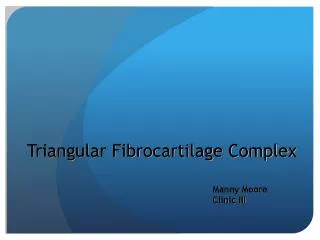
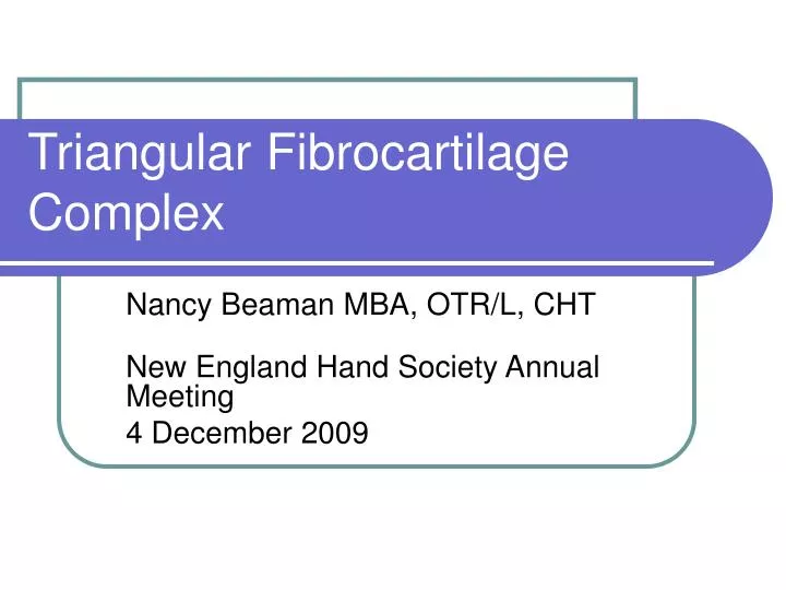
Triangular Fibrocartilage Complex. Nancy Beaman MBA, OTR/L, CHT New England Hand Society Annual Meeting 4 December 2009. TFCC: AKA…. Triangular Fibrocartilage Carpal Articular Disc Discus Articularis Triangular Ligament Triangular Cartilage Triangular Disc Meniscus Ulnocarpal Complex

E N D
Triangular Fibrocartilage Complex Nancy Beaman MBA, OTR/L, CHT New England Hand Society Annual Meeting 4 December 2009
TFCC: AKA… • Triangular Fibrocartilage • Carpal Articular Disc • Discus Articularis • Triangular Ligament • Triangular Cartilage • Triangular Disc • Meniscus • Ulnocarpal Complex • Ulnocarpal Ligament Complex
Triangular Fibrocartilage Complexa complex structure • Consists of • Triangular Fibrocartilage • Ulnocarpal ligaments
TFC Anatomy • Triangular in shape • Cartilaginous • Ligamentous • Interposed between ulnar carpus and distal ulna
Arises from articular cartilage on corner of sigmoid notch of the radius Inserts into base of ulnar styloid and volarly into ulnocarpal ligament complex Ulnocomplex: ulnolunate & ulnotriquetral ligaments TFC Anatomy
TFC Anatomy • Dorsal and volar radioulnar ligaments are fibrous thickenings w/in the dorsal and volar edges of TFC • Peripheral margin: thick lamellar collagen • Adapted to bear tensile load • Thin central portion • Articular disc: chondroid fibrocartilage • Avascular and aneural • Occasionally absent • Often so thin, is tranluscent
TFC Anatomy • Blood supply: Ant Interosseous Artery
TFC Anatomy • Radial origin 1-2mm thick at base • Stretches across ulnar articular dome • Apex attached to eccentric concavity of head and projecting styloid • Ulnar origin may be up to 5mm thick
TFC Anatomy • Volar and dorsal radioulnar ligament are confluent w/ TFC • Dorsal RU lig is stouter • Volar RU lig is origin for ulnocarpal ligs • RU ligs can be considered 2 separate laminae • Proximal lamina inserts on fovea adjacent to styloid • Distal lamina inserts into base of syloid proper • Separated by ligamentum subcruentum
Triangular Fibrocartilage The ulnocarpal or “V” ligament Combined TFC and Ulnocarpal ligaments Green’s Hand Surgery
Biomechanical Functions • Provides continuous gliding surface across distal radius and ulna for carpal flexion, extension and translation • Provides flexible mechanism for stable rotation of the radiocarpal unit about the ulnar axis
Biomechanical Functions • Suspends the ulnar carpus from the dorsal ulnar face of the radius • Connects the ulnar axis to the volar carpus • Major stabilizer of DRUJ and ulnar carpus
Biomechanical Functions • Amount of load xferred to distal ulna is directly proportional to the ulnar variance • neutral: ~20% • Positive ulnar variance • Load increases w/ corresponding decreased thickness in articular disc
(Hulten’s) Ulnar Variance • Ulnar zero: radius and ulna the same length • Ulna positive: ulna 1-5mm longer • Ulna negative: ulna 1-6mm shorter • Variance is independent of styloid length • Measurement is affected by FA rotation Grip loading Wrist position Xray techniques
Biomechanical Functions • Dynamic load xmission w/ rotation • Supination: radius moves distally creating a relative negative ulnar variance • Pronation: radius moves prox’ly creating and relative positive ulnar variance • Ulna moves w/in sigmoid notch • Dorsal with pronation • Volarly with supination • During FA rotation dorsal and volar radioulnar ligamentous portions of TFCC become tense, stabilizing DRUJ
Biomechanical Functions: Compressive Force • Force across carpal-ulnar articulation is partially transmitted thru center of TFC to ulnar dome • Tends to separate radius and ulna • Variance changes w/ grip and FA position • Neutral variance: static axial load • 80% radius, 20% ulna • w/ positive ulnar variance 2-2.5mm, load increases 5 – 40% (Palmar and Werner) • Later study by Palmer showed weak correlation
Compressive Forces • DRUJ is most stable in extremes of rotation where compressive forces are resisted by reciprocal tensile forces • Pressure concentrates • Dorsally in pronation • Palmarly in supination
Compressive Forces • In pronation • the palmar margin of TFC is taut as the dorsal margin of radius and the ulnar articular surface are compressed • If palmar margin is attenuated or torn, dorsal sublux of distal ulna occurs relative to radius
Compressive Forces • In supination • The dorsal margin becomes taut while the palmar margin of the sigmoid notch and ulnar articular surface are compressed • If dorsal margin is torn or attenuated, palmar sublux of distal ulna relative to radius occurs
Traumatic Injuries • Fall on an outstretched hand • Extension-pronation force to an axial-loaded wrist • A dorsal rotation injury • When a drill binds and rotates the wrist • A distraction force applied to the volar FA or wrist • Common with Distal Radius Fractures
Clinical History: Mechanism of InjuryTFCC injury or… • Carpal hypersupination w/ UD • ECU instab • Hyperpronation w/ dorsiflexion • Lunotriquetral ligament injury • Repeated pronation/supination • May produce chondromalacia of ulnar head • Ulnocarpal impaction, abutment or loading syndrome seen w/ ulna positive • Sudden onset of pain accompanied by redness and swelling point to inflammatory process • Frequently calicific tendonitis
Classification of Injury • Class 1: Traumatic • Central perforation • Ulnar avulsion With distal ulnar fracture Without distal ulnar fracture
Classification of Injury • Class 1: Traumatic C. Distal avulsion D. Radial avulsion With sigmoid notch fracture Without sigmoid notch fracture
Classification of Injury • Class 2: Degenerative (ulnocarpal abutment syndrome) • TFCC Wear • TFCC Wear, plus lunate &/or ulnar chondromalacia
Classification of Injury • Class 2: Degenerative (ulnocarpal abutment syndrome) C. TFCC perforation plus lunate &/or ulnar chondromalacia plus lunotriquetral ligament perforation D. TFCC perforation plus lunate &/or ulnar chondromalacia plus lunotriquetral ligament perforation plus ulnocarpal arthritis
Contribution of age • Age related changes begin as early as 3rd decade • Increase rapidly by age 50 • Virtually all wrists have significant degenerative changes and 50% or more will have arthrographically demonstrable “lesions”
Symptoms • Ulnar-sided wrist pain • Frequently with clicking • May be a history of a fall on a pronated wrist, traction or twisting injury • Improved by rest, worsened by load activity • Often assoc’d w/ tendonitis of ECU
Physical Exam • Swelling w/ a reversal of normal convex shape at ulnar border of wrist • Tenderness along soft region of ulnar border of wrist between ulnar styloid and triquetrum • Bring hand into UD compresses TFCC • RD can apply pressure to peripheral tissues • Dimpling along dorsal surface, particularly in supination—indicative of volar sublux of ulna • Ask pt to duplicate motion that causes symptoms
Physical Exam: provocative tests Only meaningful if they reproduce pt’s symptoms • TFC compression test • Examiner axially load the wrist then add UD or rotatory motion • Painful click that reproduces symptoms • Always compare to the other side • Piano key sign: test for DRUJ stability • Grasp distal ulna and attempt to passively move the ulna volarly and dorsally in various degrees of pronation and supination. • Pain and tenderness w/ inc’d motion suggest DRUJ involvement • Gross laxity represents significant TFCC disruption
Physical Exam: provocative tests • Test LT ligament w/ Ballottement test • Examiner hold lunate between thumb and IF of one hand and triquetrum in thumb and IF of the other. Move triquetrum volar and dorsal to appreciate instability or elicit pain.
Physical Exam: provocative tests • Check ECU tendon for subluxation • on supination w/ wrist-ulnar deviation, tendon displaces, often w/ audible snap, when moved in the ulnar and palmar directions • on pronation, it relocates into its normal sulcus
Physical Exam: provocative tests • Articular Disc Shear • Examiner stabilizes the distal radius while thumb is positioned dorsally over distal ulnar head • Grasp pisotriquetral complex w/ other thumb and IF • Produce a dorsal glide of pisotriquetral complex on the ulnar head producing a shearing of the articular disc • Positive if produces patient’s pain &/or laxity
Physical Exam: provocative tests • Measure grip strength • Will see decrease grip w/ TFCC injury • Pain w/ tight grip • Testing ligamentum subcruentum • In supination: pressure to distal ulna volarly should cause pain • In pronation: pressure to distal ulna dorsally should cause pain
Differential Diagnosis • ECU subluxation • LT ligament injury • Pisotriquetral arthritis • Chondral lesions of lunate or midcarpal jt • Ulnar artery thrombosis • Ulnar neuropathy at wrist
Objective testing • Differential anesthetic injection may be helpful to localize symptomatic source of pain • X-ray • Carpal alignment • Ulnar styloid morphology • Ulnar variance • MRI: to differential soft tissues • Arthrography: evaluation of integrity of intercarpal and capsular wrist ligaments • Arthroscopy
Management • Immobilization x 2 weeks (normal xray) • Reexamine after 2 weeks, continue immob if still tender • Still painful after 6 weeks then MRI • Arthroscopic evaluation • Surgical debridement • Central or peripheral tears • Repair of peripheral tear • Distal ulna resection / ulnar osteotomy
Post-operative Care: Central Debridement • Wrist Immob Orthosis and begin AROM at 3-5 days • Scar mgt once sutures removed • 3-4 weeks add resistance, PROM and dynamic splinting if needed • 6 weeks progressive strengthening
Post-operative Care Peripheral Repair • Long arm cast at 10 days when sutures removed. Short arm cast at 3 weeks. • 6 weeks: cast removed, wrist immob orthosis. Initiate AROM and scar mgt. PROM to elbow if needed. • 8 weeks: PROM & dynamic splinting if needed. • 10-12 weeks: begin strengthening.
References • Green’s Operative Hand Surgery 5th Ed • Review of Hand Surgery • Beredjiklian & Bozentka • Examination of the Hand and Wrist • Tubiana, Thomine, Mackin • Principles of Hand Surgery and Therapy • Trumble • Anatomy, Biomechanics and Pathomechanics • Handout from Drexel University, 3 Mar 07 • Atlas of the Hand Clinics: Disorders of the Lunotriquetral Joint, March 2004 • The Wrist: Diagnosis and Operative Treatment • Cooney, Linscheid, Dobyns • Rehabilitation of the Hand and Upper Extremity • Hunter, Mackin, Callahan