Download
1 / 40
410 likes | 1.1k Views
Neonatal Surgical Issues (Part 1). Sue Ann Smith, MD Neonatologist. An anatomic survey. Head and Neck lesions Chest lesions Abdomen Abdominal wall defects and infection. The Nose. Choanal atresia – bilateral atresia Respiratory distress resolves with crying

E N D
Neonatal Surgical Issues(Part 1) Sue Ann Smith, MD Neonatologist
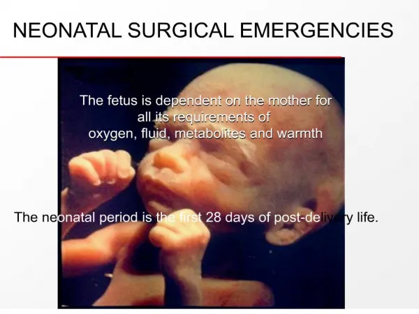
An anatomic survey • Head and Neck lesions • Chest lesions • Abdomen • Abdominal wall defects and infection
The Nose • Choanal atresia – bilateral atresia • Respiratory distress resolves with crying • Treat with oral airway until surgical repair • CT scan often used in surgical planning • ENT surgeons make opening in bony plate and stent open during healing • Nasolacrimal duct cysts – large and bilat • Respiratory distress resolves with crying • Treat with oral airway • Can usually be seen with otoscope
Robin sequence • AKA Pierre Robin syndrome • Hypoplastic mandible with U-shaped midline cleft palate • Respiratory and feeding difficulties • Position prone, may require nasopharyngeal tube, oral airway, LMA, or endotracheal tube • Mandibular distraction is now treatment of choice at OHSU
The Unusual • Laryngotracheal clefts • Laryngeal webs • Tracheal agenesis – frequently lethal • Neck masses • Foregut duplication cyst • lymphangioma
Normal Larynx Laryngeal Web
Congenital Chest Lesions • Tracheo-esophageal fistula • Diaphragmatic Hernia (briefly) • Congenital lobar emphysema • Cystic adenomatoid malformation • Vascular rings
Tracheo-Esophageal Fistula (TEF) • Esophageal atresia with TEF is most common (85%). • Diagnosis may be suspected antenatal with absence of stomach bubble and polyhydramnios. (*Caution: also seen with conditions that lead to poor swallowing) • Often associated with other anomalies: VATER and chromosomal
Tracheo-Esophageal Fistula (TEF) (cont) • Presentation: excessive salivation and intolerance of feedings. • Diagnosis: inability to pass catheter into stomach. • Pre-op Management: avoid mechanical ventilation (if possible), catheter to suction in the esophageal pouch, elevate head of bed.
Operative management • Ligation of fistula at trachea. • Mobilization of distal esophageal segment with primary anastamosis to proximal pouch. • NG tube left in place to stent open anastamosis while healing. • Chest tube left in for serous drainage usually.
Post-operative Management • Careful airway management to prevent trauma to the fistula ligation site in the trachea. • Prior to feedings, must make sure that the esophageal anastamosis does not leak. (swallow study) • Often have on going feeding problems. May need dilation procedures periodically
Other “TEFs” • Esophageal atresia without TEF – very rare • H-Type TEF-also rare. • Diagnosis usually after the neonatal period with frequent pneumonias or respiratory distress related to feedings
Congenital Diaphragmatic Hernia (CDH) • Most commonly on left side • Incidence 1:2000 to 1:5000 • Often associated with other malformations • Frequently diagnosed prenatally • Avoid bag-mask PPV
Pre-op CDH • Delayed surgical repair – usually after 72 hrs of age • NG drainage tube to keep bowel decompressed • Treat aggressively for pulmonary hypoplasia and Persistent Pulmonary Hypertension – including ECMO(?). • Surfactant therapy is now controversial
Post-Op CDH • “Anatomy is destiny” • Survival continues to be around 40-50%. • Feeding difficulties
Congenital lobar emphysema • Lesions that cause air trapping, with compression of surrounding tissue • Most common in left upper, right middle and right upper lobes • Usually attempt low volume ventilation. Sometimes selective intubation of other bronchus • May require surgical resection
Congenital Cystic Adenomatoid Malformation (CCAM) • May be confused with CDH • Abnormal lung tissue that forms fluid filled cysts. May be large cysts, or many small cysts and solid areas • Space occupying lesion • May cause shifting of mediastium • May spontaneously regress in fetus • May require surgical removal
Vascular Rings • Uncommon • Signs include stridor, vomiting and difficulty swallowing. • Barium swallow can be diagnostic, but may need chest MRI. • Sometimes may need cardiac catheterization
The Abdomen • Abdominal wall • defects • infection • Bowel • Obstructions
Gastroschisis • Abdominal wall defect to right of umbilicus with no covering over intestines • Rarely associated with other anomalies • Most babies are SGA and born to young mothers (why?) • 10% will have intestinal atresias • Rarely will have significant infarction of most of small bowel (i.e. lethal) • Most will have “meconium” stained amniotic fluid (really bile)
Gastroschisis Pre-op • Empty stomach (usually lots of bilious fluid) • NG tube for decompression • Place in bowel bag or wrap in warm saline soaked gauze and saran wrap • Support the bowel so as to maintain perfusion
Gastroschisis (post-op) • Primary closure is attempted • May require silo with slow return of intestine into small abdominal cavity • Maintain perfusion • Feeding difficulties are main post-op problem • At risk for adhesions throughout life
Omphalocele • Abdominal wall defect at umbilicus with covering (sac may rupture) • Frequently associated with other anomalies • Giant omphaloceles: respiratory issues with misshaped chest and airway malacias
Omphalocele • Decompress stomach initially • Careful eval for other anomalies • Intact sac may defer operation for years • “paint” membrane with betadine to toughen into a “rind” • Ruptured sac – repair similar to gastroschisis
Omphalitis • Presentation – erythema/induration of the periumbilical area with purulent discharge from umbilical stump. • Can spread extensively to abdominal wall or develop necrotizing fasciitis. • Both gram + and gram neg bacteria implicated • Full sepsis evaluation • Oxacillin/nafcillin and gent
Normal Larynx (upside down) Laryngeal Web (also upside down)