Download

1 / 80
900 likes | 2.6k Views
Forensic Biology Screening Workshop Semen. Semen Composition. Semen is a fluid of complex composition, produced by the male sex organs. There is a cellular component, spermatozoa, and a fluid component, seminal plasma. . Seminal Plasma.

E N D
Semen Composition • Semen is a fluid of complex composition, produced by the male sex organs. • There is a cellular component, spermatozoa, and a fluid component, seminal plasma.
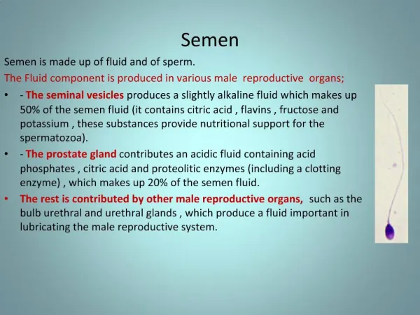
Seminal Plasma • Composed of salts, sugars, lipids, enzymes (such as Acid Phosphatase), nutrients, proteins, hormones, basic amines (spermine), P30 (Prostate Specific Antigen), flavins (source of fluorescence under UV light) • The components originate from several sources, including seminal vesicles and the prostate gland • The prostate is the source of the enzyme acid phosphatase and the protein Prostate Specific Antigen (PSA), or p30 protein
Seminal Plasma Function • Lubricates sperm duct / urethra • Nutritive and protective liquid medium for sperm to travel in • Alkaline environment which protects the sperm against the acidic nature of the vaginal tract
Seminal Plasma Vasectomy severs or ligates the ducts carrying sperm to the penis • Vasectomized men will have no sperm but will have the plasma components present in their ejaculate
Sperm Cells • Sperm are the male reproductive cells • Each consists of a head, tail and mid-piece • In humans, the head is a tiny disc, about 4.5 µm long and 2.5 µm wide • The tail is about 40 µm long, and is rapidly lost in ejaculates
Sperm Cells • The head is where the DNA is preserved • Ape sperm are similar in size and shape • Dogs have similarly shaped sperm but are about three times larger than human sperm • Other animals have differently shaped sperm
Sperm Cells • Develop in the seminiferous tubules • Spermatogenesis has three phases: • Spermatocytogenesis - mitotic division • Meiosis - diploid number present in first phase becomes haploid • Spermiogenesis – spherical sperm become elongated, acrosome forms and they are released from the seminferous tubules
Sperm Cells • Move to the epididymis • Upon physical stimulation: • Sperm and seminal fluid combine in vas deferens to form semen • Ejaculate ~ 3-4 ml of semen • ~70-150 million sperm per ejaculate
Effect On Spermatogensis • Genetic abnormalities • Disease • Injury • Chemicals – chemotherapeutic agents • Drugs • Alcohol • Age
Sperm Cell Morphology Composed of a head, midpiece and tail • Head • Acrosome cap – aids sperm entry to egg • Houses nuclear material (DNA) • ~4.5 µm long and 2.5 µm wide
Sperm Cell Morphology • Midpiece • Houses mitochondria – energy for sperm cell • Tail • Used for mobility, made of protein • Long and fragile – first part to breakdown and easily detached • ~40 µm long
Analysis of Semen • Semen stains fluorescence under ultraviolet light • It is common practice to visually assess items of evidence under UV light to locate possible semen stains • The intensity of the fluorescence can be affected by the substrate, concentration of the stain, and other body fluids
Analysis of Semen Presumptive Tests • Acid Phosphatase Detection • Human semen contains high concentrations of acid phosphatase (AP), which can therefore be the basis of a screening test. • While AP is detected in high concentrations in semen, it can also be detected in other body fluids
Analysis of Semen Confirmatory Tests • P30 Identification • Found in semen • Microscopy • Identification of sperm cells, usually done using a staining method
Analysis of Semen • Acid Phosphates degrades at a much faster rate than sperm cells • While presumptive tests for acid phosphatase can be helpful; a negative result does not necessarily mean semen is not present • Many laboratories conduct microscopic examinations on items that have negative AP test results—based on the circumstances of the case and evaluation of the item
Survival Times • Dried Stain • Sperm, Acid Phosphatase, Prostate Specific Antigen P30 – years if proper storage • Vagina / Cervix • Sperm, Acid Phosphatase = ~3 days (sperm possibly longer) • Prostate Specific Antigen P30 = ~ 1 day • Mouth • Sperm ~ 6 hours • Acid Phosphatase / Prostate Specific Antigen P30 – less due to water solubility • Anal cavity • Sperm ~ 9-20 hours • Acid Phosphatase / Prostate Specific Antigen P30 - less
Survival Times Why do sperm survive longer? • Sperm heads, very strong • Designed to last to penetrate egg • Proteins are water soluble, breakdown quicker • Sperm tails are lost first • Tails are fragile and break off • Made of proteins, bacteria attack first
Acid Phosphatase Acid Phosphatase • Secreted from the prostate • Cleaves phosphate group from molecules phosphoryl choline choline • Choline is important in cellular membrane composition and repair
Acid Phosphatase -BCIP Test HOW TO MAKE: • 0.5 mg 5-bromo-4-chloro-3-indolyl phosphate (BCIP)/ml 0.01M acetate buffer, pH 5.5 Note: Dissolve the BCIP in a few drops of dimethyl sulfoxide before bringing the solution to volume with acetate buffer HOW TO STORE: • The BCIP solution is stable for at least four weeks when stored at 4C
Acid Phosphatase -BCIP Test HOW TO PERFORM: • Swab question stain with a slightly moistened swab • Place swab into tube with 200 µl BCIP substrate solution Note: Use a known semen stain as a positive control and an unstained swab as a negative control • Incubate at 37° for 15 minutes Acid Phosphatase hydrolyzes BCIP and creates a blue color--Any blue color change is a POSITIVE result for phosphatase activity
Acid Phosphatase –AP Spot Test Reaction: • Acid Phosphatase cleaves the phosphate from α-napthyl phosphate to form napthol • Napthol and Brentamine Fast Blue B forms a purple azo dye
Acid Phosphatase – AP Spot Test HOW TO MAKE: • Buffer • Glacial acetic acid 5 ml • Sodium acetate, anhydrous (0.24M) 10 g • Distilled Water 500 ml • Solution A • Buffer 250 ml • Sodium a-naphthyl phosphate, 0.25% (w/v) 0.63 g • Solution B • Buffer 250 ml • Naphthanil diazo blue B, 0.5% (w/v) 1.25 g (Fast Blue B) Note: Both solutions can be combined for working solution in spot test or used separately
Acid Phosphatase – AP Spot Test HOW TO STORE: • If purchased from a commercial supplier, follow product insert regarding storage requirements • Solution A and Solution B can be stored frozen for months • Solution B should be stored in an amber bottle Note: when solution A and B are combined, the reagent is not as stable as when stored separately
Acid Phosphatase – AP Spot Test HOW TO PERFORM: • Take a small cutting of the question sample and place on filter paper/absorbent pad or swab the questioned evidence stain Note: Use a known semen stain as a positive control and an unstained swab or filter paper as a negative control • Add 1 drop of AP Spot Test reagent if both solutions combined or 1 drop each solution; A followed by B. • POSITIVE if purple color within 1 min. • NEGATIVE if no color change, pink color change or color change after 1 min.
Acid Phosphatase False Positives • Vaginal Acid Phosphatase • Fecal Material • Plant matter • Spermicides • Some feminine hygiene products
Acid Phosphatase (AP) Mapping Used on large items of evidence to screen for AP activity HOW TO MAKE: • Follow previous formulation, place solution A and B into separate spray bottles
AP Mapping HOW TO PERFORM: • Lay out item to be mapped onto clean surface • Staple (or otherwise attach) the appropriate size of filter paper to a piece of plastic sheeting • Spray the paper with DI water until damp. Lay flat onto item marking the position/orientation. Plastic sheeting should be on top. • Press for ten minutes (fifteen minutes if blood is mixed with semen). Use of a flat board or sheet of glass on top of sheeting can aid in this process
AP Mapping HOW TO PERFORM: • After pressing, hang paper still attached to plastic sheeting in a fume hood. • Evenly spray the paper with Solution A • Evenly spray with Solution B • Let develop for ten minutes.
AP Mapping HOW TO PERFORM: • Positive stained areas appear purple. Positive stains should appear within three minutes. Weaker stains may take longer to appear (10-15 minutes). Note: Use a known semen stain as a positive control; color reaction should occur within one minute • DO NOT overlay paper on item and mark orientation—this should have been done in first steps
AP General Swabbing Used on large items of evidence to screen for AP activity • Use a slightly moistened swab to test zones or sections of a large item • Follow the AP spot test or BCIP procedure • The technique allow the analyst to screen large items quickly
Prostate Specific Antigen (PSA) P30 Prostate Specific Antigen (P30) • Antigen made in the prostate gland • Weighs 30,000 kD • Liquefies semen and is instrumental in dissolving the cervical mucous cap for sperm entry
Prostate Specific Antigen (PSA) P30 • Early detection was based on electrophoretic or double diffusion Ouchterlony methods • The methods relied on the formation of visible precipitation of the sample and anti-p30; oftentimes followed by staining with Coomassie Blue.
P30 Electrophoresis Methods • The most widely used methods were crossover electrophoresis and rocket immunoelectrophoresis • Crossover: anit-p30 is placed into a well of an agar gel opposite of the sample and allowed to electrophoreses • The antigens and antibodies move toward each other and a precipitant formed
P30 Electrophoresis Methods • Rocket immunoelectrophoresis • a technique in which samples are placed in a row of wells in an agar plate containing antiserum (anti-P30) and an electric field perpendicular to the line of wells is applied; this drives the antigen through the gel, forming a spike or "rocket" precipitin pattern trailing away from each well. The length of the rocket is proportional to the amount of antigen placed in the well and is semi-quantitative
Elisa Test For Semen ELISA was another method introduced to aid in semen identification • Enzyme-linked immunosorbent assay (ELISA) crossover ELISA typically involves a two-stage incubated immuno reaction. First the target antigen binds with a solid phase antibody. Non-bound materials are washed away and an enzyme-labeled antibody, called a conjugate, binds to form a 'sandwich' complex. Finally the antigen-antibody is introduced to a substrate where a chromogen is used to give a color change indicating the presence of the antibody • Shown to be a very sensitive method
Commercial P30 Tests Ouchterlony, electrophoresis and ELISA methods are labor intensive--this lead to the development of faster methods • ABAcard® P30 • Seratec® PSA Semiquant • Semi-quantitative • Slight differences between the two methods
ABAcard® p30 Reaction: • “S” area – mobile monoclonal antihuman Prostate Specific Antigen P30 antibodies • Prostate Specific Antigen P30 antibodies bind to Prostate Specific Antigen P30 antigens present in sample • Form Antibody-Antigen complex • Travel through card towards the “T” area • “T” area – stationary monocolonal antihuman Prostate Specific Antigen P30 antibodies
ABAcard® p30 • Stationary antibodies in the “T” area catch the mobile Antigen-Antibody complex • Forms Antigen-Antibody -Antibody complex • Antibodies labeled with a pink dye • When antibodies aggregate – form a pink line in the “T” area
Seratec® PSA Semiquant • “C” area – internal control ensures the test card works properly. • Binds excess mobile antihuman prostate specific antigen P30 antibody • Middle line is a semi-quantitative tool (Seratec® PSA Semiquant only) • Aids in detecting semen concentration • If “T” line = middle line ~ 4ng/ml • More intense = higher concentration • Less intense = less concentration