Download
1 / 52
520 likes | 533 Views
Explore the discovery, diversity, and anatomy of cells in the human body. Learn about cell theory, sizes, structures, functions, and more.

E N D
How Many Cells are in the Human Body? • How many cells are in the human body? • Approximately 75 to 100 Trillion!
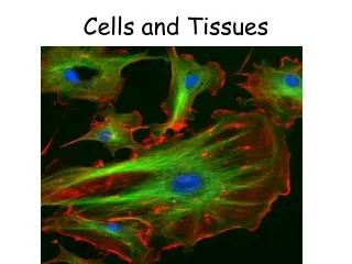
Cells • Human Physiology Chapter 3 • I. Introduction to the Cell • Definition – A cell is the smallest unit of structure and function of living things that carries on life processes.
Discovery of the Cell • Robert Hooke ( 1665) – • 1st to see cells; • looked at cork (dead plant cell); • box-shaped structures looked like monk’s rooms called ‘cells’.
Discovery of the Cell • Anton Van Leeuwenhoek (1673) – • 1st to see living cells. • Scraped mouth: cheek cells, bacteria, yeast. • Water: Protozoans, “Animalcules”.
Discovery of the Cell • Matthias Schleiden (1838) – Botanist, • concluded all plants are made of cells. • Theodor Schwann (1839) – Zoologist, • concluded all animals are made of cells • Rudolf Virchow (1855) – Physician, • concluded cells come only from other living cells.
Cell Theory • This information gave evidence for the Cell Theory: • Cells are the basic unit of structure and function in all living things. • All living things are made of cells. • Cells come only from the reproduction of other living cells
Cell Diversity • Cell Size • Most cells are only visible with microscope. • Cell size varies from 2 m to .2m (.2 x 10-6)
Cell Size • RBC • WBC • Human Egg • Smooth Muscle Cell
Cell Diversity • Examples: • Bacteria - 0.2 m (.000008 in. or 8 millioneth in.) • Giraffe leg nerve – 2 m (6 ½ ft.) • Eggs • Most body and plant cells – 10 to 50 m (.002 in.)
Cell Diversity • Why are Cells so Small?
Diffusion through the Plasma Membrane Figure 3.10
Cell Diversity • Why are Cells so Small? • Diffusion is too slow to move nutrients & wastes through cell.
Cell Diversity • Why are Cells so Small? • As cell size increases, volume increases at a faster rate than surface area. • Surface area becomes too small to allow materials to enter cell quickly enough to meet its needs.
Cell Diversity • Cell Shape • The shape of a cell helps it to perform its function.
Cell Diversity Figure 3.8a–b
Cell Diversity • Cell Shape • The shape of a cell helps it to perform its function. • Examples: • RBCs – • concave, flexible • squeeze thru vessels
Cell Diversity • Cell Shape • WBCs – • change shape • engulf particles • move to different tissues through narrow openings
Cell Diversity Figure 3.8d–e
Cell Diversity Figure 3.8f–g
Cell Diversity • Cell Shape • Nerves – • long, threadlike, branched • receive & send messages • Epithelial – • flat, packed • protection
Cell Diversity Figure 3.8c
Cell Diversity • Cell Shape • Muscles – long, rod-like, contract – pull parts together
Cells • Eukaryotes – cells that have a nucleus and membrane-bound organelles. • Prokaryotes – cells that lack a nucleus and have no membrane-bound organelles. (bacteria)
Anatomy of the Cell • Cells are not all the same • All cells share general structures • Cells are organized into three main regions: • Nucleus • Cytoplasm • Plasma membrane Figure 3.1a
The Nucleus • Control center of the cell • Directs cell activities • Three regions • Nuclear membrane • Nucleolus • Chromatin Figure 3.1b
Nuclear Membrane • Barrier of nucleus • Consists of a double phospholipid membrane • Contains nuclear pores that allow for exchange of material with the rest of the cell
Nucleoli • Nucleus contains one or more nucleoli • Lack a membrane • Dense body of RNA & Protein • Sites of ribosome production • Ribosomes then migrate to the cytoplasm through nuclear pores
Chromatin • Composed of DNA and protein • Contain genetic information • Scattered throughout the nucleus • Chromatin condenses to form chromosomes when the cell divides
Plasma Membrane • Barrier for cell contents • Double phospholipid layer • Hydrophilic heads • Hydrophobic tails • Also contains protein, cholesterol, and glycoproteins PRESS TO PLAY MEMBRANE STRUCTURE ANIMATION
Plasma Membrane Figure 3.2
Plasma Membrane Specializations • Microvilli • Finger-like projections that increase surface area for absorption Figure 3.3
Plasma Membrane Specializations • Membrane junctions • Tight junctions • Desmosomes • Gap junctions Figure 3.3
Intercellular Junctions • Tight junctions • close space between cells • located among cells that form linings • Desmosomes • form “spot welds” between • cells • located among outer skin cells • Gap junctions • tubular channels between cells • located in cardiac muscle cells
Cytoplasm • Material outside the nucleus and inside the plasma membrane • Cytosol • Fluid that suspends other elements • Organelles • Metabolic machinery of the cell • Perform specific functions to maintain life of cell • Inclusions • Non-functioning units
Cytoplasmic Organelles Figure 3.4
Cytoplasmic Organelles Mitochondrion
Cytoplasmic Organelles • Mitochondria • Double membrane, fluid-filled sacs • Folded inner membrane (cristae) • Increases surface area • “Powerhouses” of the cell • Provides ATP for cellular energy • Makes ATP from glucose • Large numbers found in muscle cells
Cytoplasmic Organelles • Ribosomes • Made of protein and RNA • Sites of protein synthesis • Found at two locations • Free in the cytoplasm • Attached to rough endoplasmic reticulum
Cytoplasmic Organelles • Endoplasmic reticulum (ER) • Interconnected membrane-bound canals • Fluid-filled ‘tubules’ for carrying substances • Intercellular Highway • Communicates with cell membrane, nuclear membrane, and organelles
Cytoplasmic Organelles • Endoplasmic reticulum (ER) con’t • Two types of ER: • Rough Endoplasmic Reticulum • Studded with ribosomes • Transports proteins • Smooth Endoplasmic Reticulum • Make lipids
Cytoplasmic Organelles • Golgi apparatus • Stack of flat membranous sacs • Modifies and packages proteins • Produces different types of packages • Secretory vesicles • Cell membrane components • Lysosomes
Golgi Apparatus Figure 3.6
Cytoplasmic Organelles • Lysosomes • Tiny membranous sacs • Contain enzymes that digest nonusable materials within the cell • Examples: • Atrophy- decrease muscle tissue during inactivity • Decrease uterus tissue after childbirth • Decrease breast tissue after weaning • Decrease tissue (‘carve out’) between fingers in fetal development
Cytoplasmic Organelles • Peroxisomes • Membranous sacs of oxidase enzymes • Detoxify harmful substances • Break down free radicals (highly reactive chemicals)
Clinical Application Diseases at the Organelle Level • MELAS – mitochondrial encephalomyopathy, lactic acidosis, and stroke-like episodes • mitochondria are missing a gene necessary to carry out important energy producing reactions • usually inherited by mother • causes strokes, severe headaches, muscle weakness and numb hands • ALD – adrenoleukodystrophy • peroxisomes are missing enzymes • causes dizziness, weakness, darkening skin, and abnormal heart rhythms • Tay-Sachs Disease • lysosomes are abnormally large and lack one enzyme • causes nervous system failure and early death
Cytoplasmic Organelles • Cytoskeleton • Network of protein structures that extend throughout the cytoplasm • Provides the cell with an internal framework • Maintains cell shape Figure 3.7a
Cytoplasmic Organelles • Cytoskeleton • Three different types • Microfilaments • Intermediate filaments • Microtubules Figure 3.7b–d
Cytoplasmic Organelles • Centrioles • Rod-shaped bodies made of microtubules • Direct formation of mitotic spindle during cell division