Download
1 / 38
560 likes | 2.62k Views
Pig Heart Dissection 101. Biology 30S. (gulp) But sheep have nice hearts…. Equipment. Pick these up (even if you are not cutting): Plastic apron Goggles Latex Gloves These should be at your table: Dissecting Tray Dissecting Kit Paper Towel Dissection Guide. Supplies you will use:.

E N D
Pig Heart Dissection 101 Biology 30S
Equipment Pick these up (even if you are not cutting): • Plastic apron • Goggles • Latex Gloves These should be at your table: • Dissecting Tray • Dissecting Kit • Paper Towel • Dissection Guide
Set Up • Start reading the dissection guide • Cut out your labels when you are finished reading (use scissors from the front) • Class orientation • Rinse your pig heart • Begin lab
Atria Ear-shaped extensions of theatria. Rt. Lt.
The atria wrap around to the anterior side of the heart. Anterior
The Coronary Vessels The cardiac muscle receives oxygen for metabolism and releases waste products into coronary vessels.
The Great Vessels The large veins & arteries, which transport blood to and from the heart muscle.
Anterior View--Aortic Arch Aorta Arising from the left ventricle of the heart is the largest artery of the body, the aorta. Rt. Lt.
Anterior View--Aortic Arch Lt. common carotid Brachiocephalic trunk (innominate artery) Rt. Lt.
Aorta: Posterior View Aorta Lt. Rt.
Posterior View--Vena Cavae Superior Vena Cava Inferior Vena Cava Lt. Rt.
Posterior View--Pulmonary Artery Lt. Pulmonary Artery Lt. Rt.
Posterior View--Pulmonary Veins Rt. Lt. Pulmonary veins are inferior to pulmonary arteries.
Rt. Pulmonary Vein Lt. Pulmonary Vein
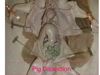
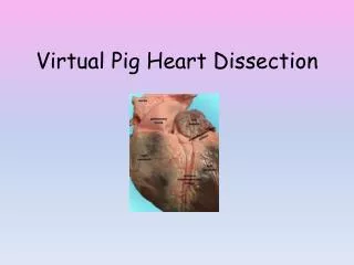
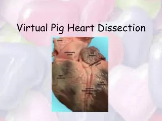
Open heart on dissection tray. Anterior half Posterior half Rt. Lt.
Rt. Atrium Lt. Atrium aorta Septum Rt. Lt.
aorta Width of right ventricular wall Width of left ventricular wall Septum Lt. Ventricle Rt. Ventricle
Tricuspid valve Cusp of valve Septum Papillary muscles Chordaetendinae
Mitral valve Septum
cusp Septum
Chordae tendinae Chordae tendinae
Check points--Exterior Heart Students should be able to point out: • Apex of heart • Ventricles & Atria • Coronary Vessels • Aorta • Pulmonary Artery & Vein • Sup & Inf Vena Cava
Check points--Interior Heart Students should be able to point out: • Septum • Atria & Ventricles • Aorta, Pulm Artery and Vein • Sup & Inf Vena Cava • Tricuspid Valve, Pulmonary Valve • MitralValve, Aortic Valve
Final Check point Students should be able to trace pathway of blood through great vessels and heart. (begin with vena cava and end with vena cava)
Anterior View • Rinse your heart under the sink and pat it dry with paper towel • Notice the coronary artery angling from the left atrium to the right ventricle
Posterior View • Label external structures
Right Side Right Side • Start your incision in the vena cava and cut through the rt. Atria and rt. Ventricle all the way to the apex.
Left Side • Start your incision in the left atrium and continue through the left ventricle to the apex.
Open heart on dissection tray. • Be sure to pat your heart dry with paper towel before attaching labels Anterior half Posterior half Rt. Lt.
That’s All folks! …and don’t forget to clean up.
Clean Up • Dispose of paper towels and labels • Hearts go into the blue plastic bag (Ms. Harder) • Wash all of your tools and rinse your dissecting trays (remove bottom and rinse if possible) • Lay removable bottoms over the edge • Place dry tools on paper towel on tray bottoms! • Leave kits open! • Wipe off all counters • Dispose of your gloves in the garbage • Wipe off your apron (pail and rag on front bench)