Download
1 / 38
380 likes | 492 Views
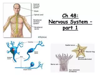
P. Ch 48 – Nervous System pt 1. Overview. Sensory input. Integration. Sensor. Motor output. Effector. Central nervous system (CNS). Peripheral nervous system (PNS). Types of neurons.

E N D
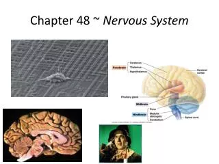
P. Ch 48 – Nervous Systempt 1
Overview Sensory input Integration Sensor Motor output Effector Central nervoussystem (CNS) Peripheral nervoussystem (PNS)
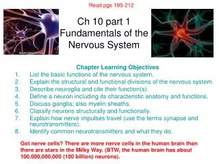
Types of neurons • Sensors detect external stimuli and internal conditions and transmit information along sensory neurons • Sensory information is sent to the brain or ganglia, where interneurons integrate the information • Motor output leaves the brain or ganglia via motor neurons, which trigger muscle or gland activity
Dendrites Parts of a neuron Stimulus Axon hillock Nucleus Cellbody Presynapticcell Axon Signaldirection Synapse Synaptic terminals Synapticterminals Postsynaptic cell Neurotransmitter
Evolutionary Adaptation of Axon Structure • The speed of an action potential increases with the axon’s diameter • In vertebrates, axons are insulated by a myelin sheath, which causes an action potential’s speed to increase • Myelin sheaths are made by glia— oligodendrocytesin the CNS and Schwann cells in the PNS
Node of Ranvier Layers of myelin Axon Schwanncell Schwanncell Nodes ofRanvier Nucleus ofSchwann cell Axon Myelin sheath
Nerve Signals • Membrane potential - the electrical charge difference across a membrane • Due to different concentrations of ions in & out of cell • Anions more concentrated inside, Cations more concentrated outside • Resting potential – the membrane potential of an unstimulated neuron • About -70 mV (more negative inside)
Changes in membrane potential act as signals • Concentration of Na+ highest outside, concentration of K+ highest inside cell. • Sodium –potassium pumps use ATP to maintain concentration gradients • Energy is potential chemical energy
A neuron at resting potential contains many open K+ channels and fewer open Na+ channels; K+ diffuses out of the cell • The resulting buildup of negative charge within the neuron is the major source of membrane potential
Key Na K Sodium-potassiumpump OUTSIDEOF CELL Potassiumchannel Sodiumchannel
Maintenance of Resting Potential - Note gradients for sodium and potassium - To maintain resting potential, you need to keep concentration gradients constant - ions have a charge, so need facilitated diffusion through membrane (is this with or against the gradient?) - there are selective ion channels that allow “leakage”
Maintenance of Resting Potential To keep sodium & potassium in right gradients, sodium-potassium pump uses ATP to maintain gradients The sodium-potassium pump pumps 2K+ in and 3Na+ out each time.
What is the result of changes in membrane potential? • Graded potentials – based on magnitude of stimulus • Action potentials – all or nothing depolarization
Types of ion channels: • Ungated ion channels – always open • Gated ion channels – open or close in response to stimuli • Ligand gated ion channels (chemically gated) ion channels–in response to binding of chemical messenger (i.e. neurotransmitter) • Voltage gatedion channels – in response to change in membrane potential • Stretch gated ion channels – in response to mechanical deformation of plasma membrane
Graded potentials • The magnitude of the change (in polarization) varies with the strength of the stimulus • These are not the nerve signals that travel along axons, but they do have an effect on the generation of nerve signals
Hyperpolarization Stimulus 50 • When gated K+ channels open, K+ diffuses out, making the inside of the cell more negative • This is hyperpolarization, an increase in magnitude of the membrane potential 0 Membrane potential (mV) Threshold 50 Restingpotential Hyperpolarizations
Depolarization Stimulus 50 • Opening other types of ion channels triggers a depolarization, a reduction in the magnitude of the membrane potential • For example, depolarization occurs if gated Na+ channels open and Na+ diffuses into the cell 0 Membrane potential (mV) Threshold 50 Restingpotential Depolarizations 100 0 3 4 5 1 2
Graded potentials occur up to a particular voltage, the threshold voltage • If depolarization reaches the threshold voltage, then an action potential is triggered.
Action Potentials • Signals conducted by axons, transmitted over long distances • Occur as the result of gated ion channels that open or close in response to stimuli • A specific dynamic change in the charge across the membrane of a cell, one that occurs either totally or not at all (“all or nothing”
Action Potential 50 Actionpotential • Steps: • 1) resting state • 2) threshold • 3) depolarization phase • 4) repolarization phase • 5) undershoot 0 Membrane potential (mV) Threshold 50 Restingpotential 100 0 3 5 6 4 1 2
Resting state • Most voltage gated sodium(Na+) and potassium gated (K+) ion channels are closed, membrane potential is at -70 mV
Threshold An electric stimulus causes the voltage-gated Na+ channels open first and Na+ flows into the cell The flow of Na+ causes more voltage – gated Na+ channels to open, so more depolarization (what kind of feedback is this) If this depolarization reaches threshold potential, then more Na+ gates open
Depolarization phase • Membrane potential rises rapidly as sodium ions rush into cell • Also known as “rising” phase
Repolarization phase • The Na+ voltage gates close shortly after opening, so Na+ inflow stops • The K+ voltage gates open, so K+ rushes out • There is a loss of positive charge, so cell returns to resting state • “Falling” phase
Undershoot • The membranes permeability to K+ is higher than at rest, so membrane potential dips down lower than resting potential • K+ gates close, so membrane potential returns to normal
Refractory period • After an action potential occurs, there is a period of time when another action potential cannot be triggered • During undershoot – sodium channel inactivation gates are closed – haven’t had enough time to reopen yet
1 5 4 3 2 1 5 4 3 1 2 Key Na K Falling phase of the action potential Rising phase of the action potential 50 Actionpotential 0 Membrane potential(mV) Threshold 50 Depolarization Resting potential 100 Time OUTSIDE OF CELL Sodiumchannel Potassiumchannel INSIDE OF CELL Inactivation loop Resting state Undershoot
How do action potentials “travel” along a neuron? • Where action potential is generated (usually axon hillock), the electrical current depolarizes the neighboring region of membrane • Action potentials travel in one direction – towards synaptic terminals
1 Axon Plasma membrane Actionpotential Cytosol Na Action potentials are self-propagating across the axon.
2 1 Axon Plasma membrane Actionpotential Cytosol Na Actionpotential K Na K
2 1 3 Axon Plasma membrane Actionpotential Cytosol Na Actionpotential K Na K Actionpotential K Na
Why doesn’t it travel backwards? • The refractory period is due to inactivated • Na+ channels, so the the depolarization can only occur in the forward direction.
Speed of action potentials • Speed is proportional to diameter of axon, the larger the diameter, the faster the speed • Several cm/sec – thin axons • 100 m/sec in giant axons of invertebrates such as squid and lobsters
Nerveswith giant axons Ganglia Brain Arm Eye Mantle Nerve http://www.youtube.com/watch?v=omXS1bjYLMI
Speeding up Action potential in vertebrates Schwann cell • Myelination (insulating layers of membranes) around axon • Myelin is deposited by Schwann cells or oligodendrocytes. Depolarized region(node of Ranvier) Cell body Axon
Action potentials are formed only at nodes of Ranvier, gaps in the myelin sheath where voltage-gated Na+ channels are found • Action potentials in myelinated axons jump between the nodes of Ranvier in a process called saltatoryconduction