Download
1 / 17
190 likes | 738 Views
History of Neuroanatomy. Neuron Doctrine Rudolf Albert von Kölliker (1817-1905) Heinrich Wilhelm Gottfried Waldeyer (1837-1921) Camilio Golgi (1843-1926) Santiago Ramon y Cajal (1852-1934). 19th Century Neuroanatomy. Rudolf Albert von Kölliker (1817-1905)

E N D
History of Neuroanatomy Neuron Doctrine Rudolf Albert von Kölliker (1817-1905) Heinrich Wilhelm Gottfried Waldeyer (1837-1921) Camilio Golgi (1843-1926) Santiago Ramon y Cajal (1852-1934)
19th Century Neuroanatomy Rudolf Albert von Kölliker (1817-1905) nucleus of Kölliker (lamina X of Rexed), naming “axon” Proponent of neuron doctrine
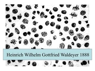
19th Century Neuroanatomy Heinrich Wilhelm Gottfried Waldeyer (1837-1921) Waldeyer cell of spinal dorsal horn, Proponent of neuron doctrine
19th Century Neuroanatomy Camilio Golgi (1843-1926) Golgi method (Golgi’s metallic impregnation) Golgi type I & II cell Golgi apparatus Golgi tendon organ Golgi-Mazzoni corpuscle
19th Century Neuroanatomy Santiago Ramon y Cajal (1852-1934) Neuron doctrine Cajal's gold-sublimate method for astrocyte horizontal cell of Cajal (Retzius-Cajal cell) of cerebral cortex interstitial nucleus of Cajal
20th Century Neuroanatomy Neuroanatomy of the 20th Century Rapid Explosion of Neuroscience 1990s the "Decade of the Brain". 31 NobelLaureates in Physiology and Medicine (16 year) Renaissance of the Neuroanatomical Techniques
20th Century Neuroanatomy Renaissance of the Neuroanatomical Techniques Neural Tissue Culture Electron Microscopy Silver Impregnation for degenerating fibers Autoradiographic tracing of neural pathways HRP tracing of neural pathways Single cell staining technique Fluorecence Histochemistry Immunohistochemistry In situ Hybridization Neuroimaging techniques (CT, MRI, PET )
20th Century Neuroanatomy Neural Tissue Culture (1907) Ross Granville Harrison (1870-1959)
20th Century Neuroanatomy Electron Microscopy 1931 Ruska and colleagues built the first electron microscope 1939 Siemens - first commercial EM 1952 Palade, Porter, and Sjöstrand developed fixation and thin sectioning
20th Century Neuroanatomy Sanford L. Palay (b. 1918) • first to visualize • synapse by • electron microscopy
20th Century Neuroanatomy Tract Tracing Studies Silver Impregnation for degenerating fibers Glees (1946) Nauta (1950) Nauta-Gygax (1954) Fink-Heimer (1967) Autoradiographic tracing of Neural Pathways Grafstein (1967) HRP tracing of Neural Pathways Kristensson & Olsson (1971) LaVail & LaVail (1972)
20th Century Neuroanatomy Single Cell Staining Technique Procion Yellow - Stretton & Gravitz (1968) Cobalt Chloride - Tweedle & Cohen (1972) HRP - Jankovska, Radstad & Westman (1976); Snow, Rose and Brown (1976)
20th Century Neuroanatomy Fluorecence Histochemistry Heimstädt (1911) constructed fluorescence microscope Stübel (1911) first uses fluorescence microscope Max Haitinger (1930s) popularized FM Albert Coons (1941): beginning of immunofluoresence Farck-Hillarp method (1962) for cellular localization of monoamines Dahlström and Fuxe (1964) applied Falck-Hillarp method in CNS
20th Century Neuroanatomy Immunohistochemistry 1941 Coons and colleagues developed indirect immunofluorescence method 1966 Graham & Karnovsky, developed enzyme (horseradish peroxidase) tagging method 1970 Sternberger developed peroxidase-antiperoxidase(PAP) technique 1971 Hökfelt applied immunohistochemistry to visualize neurotransmitters
20th Century Neuroanatomy In situ Hybridization 1969 Pardue and Gall first develops in situ hybridization technique 1983 Gee and colleagues applied in situ hybridization technique in the CNS 1986 Young and colleagues popularized quantitative in situ hybridization in nervous tissue
20th Century Neuroanatomy Neuroimaging Techniques CT (Computed Tomography) Hounsfield (1972) PET (Positron Emission Tomography) Phleps, Hoffman, and Ter Pogossian (1974) MRI (Magnetic Resonance Imaging) Lautebur (1972) Mansfield and Grannell (1973) 1979 first commercial MRI
References Clarke E, Dewhurst K: An Illustrated History of Brain Fuction. Imaging the Brain from Antiquity to the Present. 2nd ed., San Francisco, Norman, 1996 Clarke E, O'Malley CD: The Human Brain and Spinal Cord. A Historical Study Illustrated by Writings from Antiquity to the Twentieth Century. Berkley, University of California, 1968 Corsi P (ed): The Enchanted Loom : Chapters in the History of Neuroscience. New York, Oxford University, 1991 Finger S: Origins of Neuroscience. A History of Explorations into Brain Function. New York, Oxford University, 1994 Marshall LH, Magoun W (eds): Discoveries in the Human Brain: Neuroscience Prehistory, Brain Structure, and Function. Totowa, Humana, 1998 McHenry LC: Garrison's History of Neurology. Springfield, Charles C. Thomas, 1969 Meyer A: Historical Aspects of Cerebral Anatomy. London, Oxford University, 1971 Samson F, Adelman G (eds): The Neurosciences. Paths of Discovery, II., Boston, Birkhauser, 1992 Worden FG, Swazey JP, Adelman G (eds): The Neurosciences. Paths of Discovery, I., Boston, Birkhauser, 1992