Download
1 / 93
930 likes | 952 Views
Learn about the pectoral girdle and upper limbs, including the bones of the shoulder, arm, forearm, wrist, palm, and fingers. Discover their surface markings and common disorders.

E N D
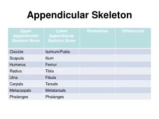
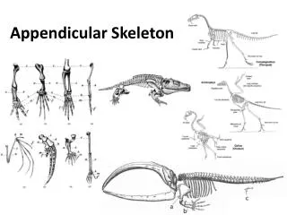
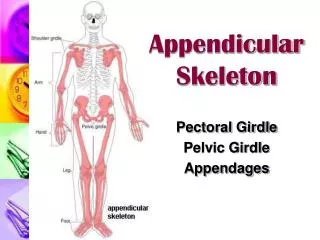
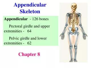
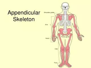
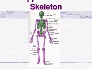
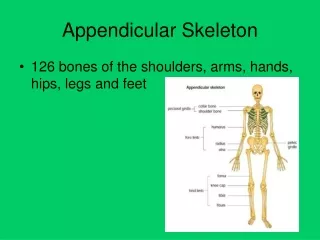
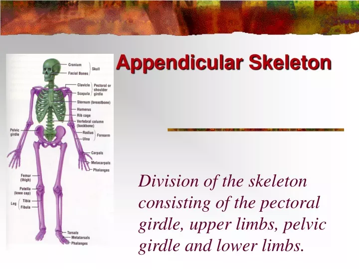
Appendicular Skeleton Division of the skeleton consisting of the pectoral girdle, upper limbs, pelvic girdle and lower limbs.
Pectoral Girdle • Attaches the bones of the upper limbs to the axial skeleton
Clavicle • Also known as the collarbone • Long, slender S-shaped bone that lies horizontally above the first rib (Transmits mechanical force from the upper limb to the trunk)
Scapula • Also known as the shoulder blade • Large, flat triangular bone on the posterior part of the thorax
SPINE: A sharp ridge that runs diagonally across the back portion of the scapula body • BODY – Main flat area of the scapula • ACROMION: The lateral end of the spine. Where the scapula articulates with the clavicle
GLENOID CAVITY (glenoid fossa) - a depression inferior to the acromion where the head of the humerus sits • CORACOID PROCESS – Projection anterior to the acromion for muscle attachment
Upper Limb • Consists of 30 bones (all paired up) • Humerus in the arm • Ulna and radius in the forearm • 8 carpals, 5 metacarpals, and 14 phalanges in the hand
Humerus • Longest and largest bone of the upper limb • Articulates with the scapula at the shoulder and both the ulna and radius at the elbow
Humerus Bone Surface Markings • ANATOMICAL NECK: constricted portion distal to the head – site of the epiphyseal plate
BODY: Main portion of the bone (diaphysis) • DELTOID TUBEROSITY: a roughened V-shaped area where the deltoid muscle attaches
CAPITULUM – small rounded process at the distal end that articulates with the head of the radius. • RADIAL FOSSA - a depression that receives the head of the radius when the forearm is bent.
TROCHLEA - a spool-shaped surface that articulates with the ulna. • CORONOID FOSSA – a depression that receives part of the ulna when the forearm is bent. • OLECRANON FOSSA - a depression on the back of the bone that receives the ulna when the forearm is straightened.
Ulna • Located on the medial side of the forearm (pinky side) • Longer than the radius
Ulna Bone Surface Markings • The olecranon forms the prominence of the elbow on the proximal end. • The coronoid process projection on the proximal, helps to hold the trochlea • Trochlear Notch – depression formed by the olecranon and coronoid process
The radial notch is a depression for the head of the radius. • A styloid process is a pointy projection at the distal end.
Radius • Located on the lateral side of the forearm (thumb side)
Radius Bone Surface Markings • Radial tuberosity a raised, roughened area that is where the biceps brachii muscle attaches to the bone • Styloid Process – pointy projection on the distal end
Carpus (Wrist) • 8 carpals • Held together by ligaments with four bones in each row • Named for their shapes • Short bones
The carpals in the proximal (closest to the radius/ulna) row are the: • Scaphoid, Lunate, Triquetrum, and Pisiform • The carpals in the distal row are the: • Trapezium, Trapezoid, Capitate, and Hamate
Metacarpus (Palm) • 5 metacarpals • Each consists of a proximal base, an intermediate body, and a distal head • Numbered I-V starting with the thumb • Long bones
Phalanges (Fingers) • 14 in each hand • Thumb has two (proximal and distal) • In each of the other four digits, there are three (proximal, middle, and distal)
Carpal Tunnel Syndrome • Narrowing of the carpal tunnel causes compression of the median nerve • The nerve compression causes pain, numbness, tingling, and hand muscle weakness
Rotator Cuff Injury • Tears or inflammation of ligaments and tendons of the shoulder near the humerus • Results in pain and loss of shoulder mobility
Checkpoint Questions • Which bones make up a pectoral girdle? What is the function of the pectoral girdle? • With which part of the scapula does the humerus articulate? • What part of the ulna is called the “elbow”? • What part of which bones are commonly called the “knuckles”? • What bones form the upper limb, from proximal to distal?
Pelvic (hip) Girdle • Functions: • Support for vertebral column • Protect pelvic organs • Attach lower limbs • Coxal Bones: Hip bones • 3 parts: Pubis, Ilium and Ischium
Articulations • Sacroiliac Joint – posterior articulation of the pelvic girdle • Pubic Symphysis – anterior articulation of the pelvic girdle • Acetabulum – attachment point of the femur • socket of the ball and socket joint
Coxal Bones • Pubis – anterior portion • Joined by pubic symphysis • Ilium – superior portion • Iliac Crest – ridge at the top of the ilium • Ischium – inferior portion • Acetabulum – socket for the head of the femur • Obturator Foramen – hole formed by the ischium and pubis
Pelvis • Combination of the sacrum, coccyx, and the 2 hip bones • Greater (false) – Top portion that is not fully enclosed by bone • Lesser (true) – Bottom portion the is completely surrounded by bone
Pelvis - continued • Ilium • Ischium • Pubis • Pubic Symphysis • Sacrum • Coccyx • Pelvic Brim • Pubic Arch
Pelvimetry • Measurement of the size of the inlet and outlet of the birth canal.
Pelvic Girdle Checkpoint • What is the name of the hip bone? • What are the 3 parts of the hip bone? • Can you identify them on a diagram??? • List 3 functions of the pelvic girdle • What is the name of the “socket” where the head of the femur sits? • List 3 differences between the male and female pelvis. Why are these present?
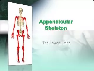
LOWER LIMB • Includes the thigh, leg, ankle, foot and toes • 30 bones in each • Femur • Patella • Tibia • Fibula • Tarsals • Metatarsals • Phalanges
Femur • Thigh Bone • Longest, strongest heaviest bone in the body • Diaphysis has a medial bend to bring knees closer to the midline of the body
Femur continued • Body - diaphysis • Head – “ball” of ball and socket joint • Neck – common site of fractures • Greater and Lesser Trochanters – used for muscle attachment • Lateral and Medial Condyles – articulation with the tibia • Patellar Surface
Patella • Sesamoid bone • Develops in the tendon of the quadriceps femoris muscle • Increases the leverage of the tendon and maintains the position of the tendon
Patellofemoral stress syndrome • AKA “Runner’s Knee” • Patella does not glide up and down between the femoral condyles but rather laterally causing pain.