Download
1 / 34
350 likes | 652 Views
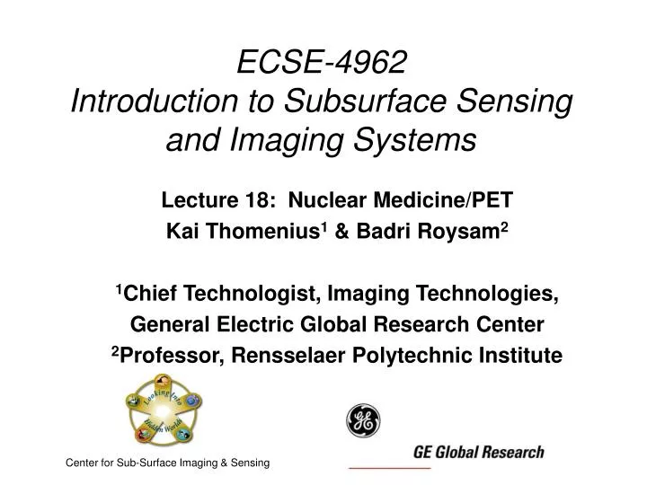
ECSE-4962 Introduction to Subsurface Sensing and Imaging Systems. Lecture 18: Nuclear Medicine/PET Kai Thomenius 1 & Badri Roysam 2 1 Chief Technologist, Imaging Technologies, General Electric Global Research Center 2 Professor, Rensselaer Polytechnic Institute.

E N D
ECSE-4962Introduction to Subsurface Sensing and Imaging Systems Lecture 18: Nuclear Medicine/PET Kai Thomenius1 & Badri Roysam2 1Chief Technologist, Imaging Technologies, General Electric Global Research Center 2Professor, Rensselaer Polytechnic Institute Center for Sub-Surface Imaging & Sensing
Recap • Phase information adds much value to images • Not just Beer-Lambert anymore! • Spectral response provides substance-specificity to imaging • Fluorescence and Multi-photon imaging are powerful tools which supply biology-specific info. • QTM is a highly sophisticated imaging method which makes heavy use of phase information. • Optical techniques have great success with near surface imaging. • How can we take this deeper from the surface? • That’s today’s topic. • We will be talking about means for detecting, not fluorescence, but signals from radioactive sources.
Nuclear Medicine/PET • For the most part in this course, our focus has been on imaging physical objects. • We have looked for features which interact with our probes • Attenuation with X-ray • Impedance mismatches in pulse-echo methods • Variations in proton density in MRI (next) • Nuclear medicine & PET are quite different • As with fluorescence-based methods, they image concentrations of exogenous chemicals injected into the patient • The observability of these is often based on radioactivity.
Nuclear Medicine • Imaging is done by tracing the distribution of radiopharmaceuticals within the body. • Radionuclides or radioisotopes are atoms that undergo radioactive decay and emit radiation. • In nuclear medicine, we are interested in radionuclides that emit x-rays or gamma rays. • A radiopharmaceutical is a radionuclide bound to a biological agent. • The role of the biological agent is the key: it gives us clinical specificity. Siemens Gamma Camera
Physics of Nuclear Medicine • 3 basic mechanisms for photon - matter interaction: • Photoelectric Effect • Compton Scatter • Pair Production • Any one of these can happen to the gamma-rays which emanate from the radionuclides. Compton Scatter Pair Production
Energy of a Gamma Ray • Radionuclide has a typical energy: e.g. 140 keV for 99mTc • Detection of lower energy scattered gamma- or x-rays degrades contrast and image quality. • A radioisotope emits one (or more) very sharp energy lines
How does this work? • Radioisotopes are injected into the body • A radioisotope can be: • a pure element (e.g. I-131 which connects to Thyroid) • a biological agent labeled with radioisotopes like MIBI-Tc99m • All isotopes have a half life. • All isotopes are expelled from the body with an associated half life. • Nuclear Medicine provides physiological images, i.e. the metabolic activity of the organs process the radiopharmaceutical and concentrate it in the target organs for imaging.
Detector or Scintillator • (NaI): Emits light whenever hit by gamma ray. Amount of light is proportional to gamma energy level. • Photomultiplier Tubes: read the light signals and translate them into electrical signals
Cross-section of an Anger Camera • Shield Around Head • Mounting Ring • Collimator Core • Sodium Iodide Crystal • Photomultiplier Tubes Gamma Camera invented by Harold Anger in early 50s.
Nuclear Medicine Performance Metrics • Typical performance: • Energy resolution: 9.5 – 10% • FWHM response • Spatial resolution: 3.2 – 3.8 mm • Uniformity: 2 – 4%
Collimator Design & Function Resolution v. Efficiency Trade-off
Nuclear Medicine Images • Typical image: • 64 by 64 pixels • Intensity gives “counts per pixel” • Pseudocolor often used. • Nuclear med imaging modes: • Static • Dynamic • MUGA (multi-gated acquisition) • Whole Body • SPECT
Cardiac Study • Evaluation of the coronary artery circulation • Myocardial perfusion • 3D Studies of the radionuclide activity
SPECT Scanners • Single Photon Emission Computerized Tomography • Store radionuclide emission data from multiple projections • Projections taken every 3 or 6 degrees. • Use CT type algorithms to determine the location and degree of accumulation of agent.
PET Imaging • What is PET Imaging? • A technique tracks biochemical and physiological processes in vivo • Uses tracer compounds labeled with positron-emitting radionuclides • As such, it is considered a form of functional imaging • Positron Emission Tomography • Functional v. Anatomical Imaging: Choose: • X-Ray - Nuclear Medicine • MRI - CT • US • How is PET different from Nuclear?
PET – Positron Emission Tomography • Certain radionuclides emit positrons. • When a positron meets an electron, they annihilate each other. • This annihilation results in a generation of two gamma rays. • The gamma rays travel in opposite directions. • The energy of these gamma rays is 511 KeV. • PET Imaging is based on detection of these gamma rays.
PET Systems Event Detection • Several gamma-detector rings surround the patient. • When one of these detects a photon, a detector opposite to it, looks for a match. • Time window for the search is few nanosecs. • If such a coincidence is detected, a line is drawn between the detectors. • When done, there will be areas of overlapping lines indicating regions of radioactivity. LOR – Line of Response
Coincidence Events • Three Types: • True • The event we are after • Scatter • At least one Compton scatter event • Wrong LOR • Random • Unlucky break • Current hot topic: • Time-of-flight PET • Estimate the arrival time at the two detectors. • Picosecond electronics
How Does PET Compare With Other Imaging Modalities? • PET provides images of molecular-level physiology • accumulation of the metabolically active radionuclide • Extends capabilities of other modalities. • Like CT, it uses tomographic algorithms • Like Nuclear, images represent distributions of radiotracers. • That’s where the similarity ends. Here are three studies of a patient done with CT, MRI, and PET scanners. CT Scan MRI Scan PET Scan Report: Patient Deceased. Report: Normal Report: Normal
PET Scanner Components • Scanner - to perform the clinical exam • Includes a tomographic reconstruction • Cyclotron - to produce the positron-emitter • Tracer Lab Equipment - to produce the tracer http://laxmi.nuc.ucla.edu:8000/lpp/radioisotopes/radioisoprod.html#CycloOp
PET Radiotracer Evolution • Initially 15O ion labeled to O2, CO, and CO2 • Main applications in brain oxygen • In mid 70s, cyclotrons were brought in. • FDG, a glucose based tracer introduced. • FDG is now the dominant PET tracer
PET Radiotracers • 18FDG is probably the most widely used PET tracer. • Fluorodeoxyglucose • Glucose based, hence high metabolic relevance • High FDG pick-up by tumors first reported in 1980 at Brookhaven NL. • Can also be used to measure rate of metabolism in brain.
Application in Lung Cancer • Case Study: • 55-year old female • Lung Cancer • 2 cycles of chemo & radiotherapy • PET results: • Increased uptake of FDG in lung nodules • Increased uptake of FDG in lymph nodes • Conclusion: Therapy will have to be continued.
PET/CT Scanners • Generation of PET & CT images in a single study • The image data sets are registered and fused. • Anatomic data from CT • Metabolic data from PET • Colorectal Cancer shown in images.
・ 56 year old male ・ Patient complained of left scapular pain ・ Chemotherapy courses performed for primary cancer of unknown origin ・ Post-therapy PET/CT scan reveals gastro-esophageal cancer with distal esophageal 18F-FDG uptake ・ Additional left scapular bone FDG uptake ・ Image courtesy of Medical College of Wisconsin, USAhttp://www.gemedicalsystemseurope.com/euen/rad/nm_pet/products/pet_sys/discovery_st_ig.html CT Scan PET Scan Fused Images PET/CT Imaging
PET & Molecular Imaging (MI) • There is a strong similarity w. PET & MI. • PET is often classified under MI. • There is a significant distinction, however. • MI probes are often designed to interact w. cellular processes. • This interaction is used to improve detectability. • PET probes are usually passive in this regard. • They rely on the inherent radioactivity of the probes.
Source Material • http://apps.gemedicalsystems.com/geCommunity/nmpet/nmpet_neighborhood.jsp • Siemens & Philips web sites for nuclear medicine & PET • http://www.crump.ucla.edu/software/lpp/lpphome.html • http://thayer.dartmouth.edu/~bpogue/ENGG167/13%20Nuclear%20Medicine.pdf • http://zoot.radiology.wisc.edu/~block/bme530lectures/L01Intro.ppt
Summary • Introduction to Nuclear Medicine and PET imaging. • Additional examples of agents (probes) introduced to reveal subsurface phenomena. • Today’s focus on radioactive labeling. • Review of instruments • Relatively straightforward devices. • Signal-to-noise ratio challenges, need to limit exposure. • Powerful clinical tools. • Much of today’s research focused on PET and extensions of PET technology.
Homework: Lecture 25 • Using internet sources, • discuss the patient and clinician safety issues from the use of radioactive tracers in PET and nuclear imaging. • SPECT imaging is a form of the scanners we discussed today. Review its theory of operation and discuss how SPECT imagers use the computed tomography algorithms (e.g. filtered backprojection).
Instructor Contact Information Badri Roysam Professor of Electrical, Computer, & Systems Engineering Office: JEC 7010 Rensselaer Polytechnic Institute 110, 8th Street, Troy, New York 12180 Phone: (518) 276-8067 Fax: (518) 276-6261/2433 Email: roysam@ecse.rpi.edu Website: http://www.rpi.edu/~roysab NetMeeting ID (for off-campus students): 128.113.61.80 Secretary: Laraine Michaelides, JEC 7012, (518) 276 –8525, michal@rpi.edu
Instructor Contact Information Kai E Thomenius Chief Technologist, Ultrasound & Biomedical Office: KW-C300A GE Global Research Imaging Technologies Niskayuna, New York 12309 Phone: (518) 387-7233 Fax: (518) 387-6170 Email: thomeniu@crd.ge.com, thomenius@ecse.rpi.edu Secretary: Laraine Michaelides, JEC 7012, (518) 276 –8525, michal@rpi.edu