Download
1 / 69
700 likes | 984 Views
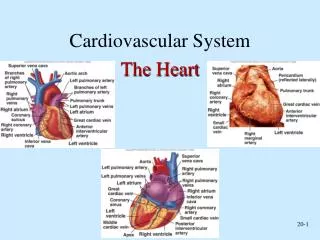
Cardiovascular system and Heart ischemia (infarction) incl. Detection of heart ischemia using bioimpedance measurement Andres Kink 2012. C ONTENTS C ARDIOVASCULAR SYSTEM M YOCARDIAL ISCHEMIA A NATOMY OF THE HEART C ARDIOVASCULAR SUSTEM AND CORONARY CIRCULATION

E N D
Cardiovascular system and Heart ischemia (infarction) incl. Detection of heart ischemia using bioimpedance measurement Andres Kink 2012
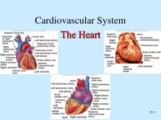
CONTENTS CARDIOVASCULAR SYSTEM MYOCARDIAL ISCHEMIA ANATOMY OF THE HEART CARDIOVASCULAR SUSTEM AND CORONARY CIRCULATION CARDIAC RHYTM AND ARTIFICIAL PACING PRINCIPLES OF RATE CONTROL
CARDIOVASCULAR SYSTEM Energy as product of low temperature burning of food products inside the body To maintain life, every living animal organism must have additional energy inflow as food and oxygen. To save excess of food for future is possible due to intracellular systems. But the same is not possible for oxygen. Oxygen is gaseous, and to accumulate it inside the body in reasonable quantity will take too much energy. In this text we will focus on energy as energy units (joule) or as units of used oxygen to get energy.
CARDIOVASCULAR SYSTEM Oxygen and food substrate delivery system for cells Most of animals (not included fishes) have specialized oxygen carrying system to maintain body tissues oxygenation: • Blood as solute to carry oxygen • Lungs as barrier between atmosphere and blood. • Circulation system as tubing system to carry oxygen rich blood to every cell in body, and to collect waste from it. • Cellular system to produce ATP from energetic substances and oxygen.
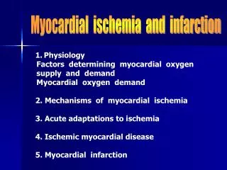
MYOCARDIAL ISCHEMIA Definition: Myocardial ischemia is an imbalance between oxygen supplyof the myocard and oxygen demand of the myocard. In general ischemia is a decrease in the blood supply to a bodily organ, tissue, or part caused by constriction or obstructionof the blood vessels. In the case of the heart the ischemia means a narrowing of the coronary artery(s) sufficient to prevent adequate blood supply to the myocardium. This narrowing may progress to a point where the heart muscle is damaged (infarction). _____________________________________ MYΣ+KAPΔΊA = myocard (muscle + heart, in the contemp. Greek: ο μυς της καρδιάς) (in Latin: MUS(CULUS) = mouse, muscle) IΣX…+AΊMIA = isch(a)emia (Greek: stop+blood) IN+FARCTUS = infarct (Latin: in+filled)
MYOCARDIAL ISCHEMIA Possible types of ishemia
MYOCARDIAL ISCHEMIA Ishemia as energy imbalance • Energy imbalance is result of non-equal oxygen supply related to oxygen consumption. • Ischemia with myocardial cell damage is often described in heart as myocardial infarction.Myocardial infarction is not reversible process, cell necrosis is healed by scar formation. • Short time myocardial ischemia is not dangerous, because myocardial has limited protection against lack of oxygen.
MYOCARDIAL ISCHEMIA Epidemiology • Heart ischemic conditions are most leading reason for mortality in world. • Silent myocardial ischemia is dangerous condition witch leads very offen to myocardial infarction (muscle tissue necrosis)
MYOCARDIAL ISCHEMIA Ischaemic heart disease world mapDALY - WHO2004
CARDIOVASCULAR SYSTEM, and CORONARY CIRCULATION Physiology of the coronary arteries
CARDIOVASCULAR SYSTEM, and CORONARY CIRCULATION Coronary artery disease
CARDIOVASCULAR SYSTEM, and CORONARY CIRCULATION Coronary reserve
CARDIOVASCULAR SYSTEM, and CORONARY CIRCULATION Special Features of Coronary Circulation • At rest, coronary blood flow BF = 5% of cardiac output CO = 250ml/min = 60-80ml/100gm/min • During exercise rises by 2 … 5 times (coronary vasculature has a high vasodilator reserve capacity) • Coronary Blood Flow is phasic • Total Coronary Flow is greater during diastole Therefore, the most crucial factors for perfusing coronary arteries are - aortic pressure - duration of diastole
CARDIOVASCULAR SYSTEM, and CORONARY CIRCULATION Myocardial O2 demand • The cardiac muscle always depends on aerobic oxidation of substrates, even during heavy exercising • The cardiac muscle has the highest O2 uptake (VO2) compared to other tissues of the body (12…15 volume%; 7…9 ml O2/100gm/min) • This is achieved by a dense network of capillaries, all is perfused at rest (no capillary reserve) • Maximal extraction of O2 from RBCs (almost no reserve of O2 extraction)
CARDIOVASCULAR SYSTEM, and CORONARY CIRCULATION Pressure volume area inside of ventricles (left ventricle)
CARDIOVASCULAR SYSTEM, and CORONARY CIRCULATION Cardiac cycle and pressure-volume area Cardiac Output (CO) determined thru Heart Rate (HR) and Stroke Volume (SV)
CARDIOVASCULAR SYSTEM, and CORONARY CIRCULATION Frank Starling Curves • Ability of the heart to change force of contraction in response to changes in venous return. • If EDV increases, there is a corresponding increase in stroke volume, suggesting heart failure and inotropy. • Reduced stroke volume suggests increased preload and decreased ejection fraction.
CARDIOVASCULAR SYSTEM, and CORONARY CIRCULATION Cardiac Output • Cardiac Output is the volume of blood (in liters) ejected by the heart in one minute • Stroke Volume is the volume of blood (in liters) ejected by the heart in one beat • When the body is under stress (physical, emotional), the heart tries to increase cardiac output … by increasing the rate according to this formula Cardiac Output = Heart Rate xStroke Volume CO = HR x SV
CARDIOVASCULAR SYSTEM, and CORONARY CIRCULATION Bradycardia or “low heart rate”
Artifical heart assisting devices The first artificial pacemaker to maintain heart rhythm was induced by Steiner in Germany to avoid cardiac arrest as a side effect of chloroform anaesthesia. Steiner's study (1871) was performed in chloroform arrested hearts of horses, donkey, dogs, cats and rabbits. In the next year the same method was used in humans by Green in the United Kingdom. The first pacemakers had interrupted galvanic (direct-current) stimuli and were connected by 13 cm long needles directly to the myocardium.
Modern era of implantable pacemakers • The first implantable pacemaker was made by Swedish inventor Dr. Rune Elmqvist, and implanted in 1958 by Dr. Ake Senning. • The first demand pacemaker was introduced by Berkovits in June 1964. Demand pacemaker have additional sensing unit to avoid competition with heart own pacemaker (sinus node), and to save battery energy.
Sensory systems • ECG based interval measurements • Movement analysis (acceleration, ..) • Temperature measurement • Impedance based • Lung impedance • Intraventricular impedance, mostly right ventricle • Myocardial impedance
Rate adaptive pacing • Heart rate is regulated to maintain body energetic needs • First generation target was night time heart rate reduction • New generation is multisensor (accelometer, ECG, temperature, bioimpedance based, …) based optimal heart rate calculation
Why Rate Response? • Rate response is the pacemaker’s ability to increase the pacing rate in response to physical activity or metabolic demand • Rate response mimics the healthy heart • Special sensor(s) required • Accelerometer • Piezoelectric crystal • Minute ventilation (transthoracic impedance) • Blood temperature • Single or combination
Chronotropic Incompetence If the patient’s heart cannot increase its rate appropriately in response to increased activity, the patient is chronotropically incompetent Chronotropic incompetence (definitions): • Maximum heart rate < 90% x (220 - Age) • Maximum heart rate < 120 bpm Causes • aging • drugs • heart disease
Sensors • Rate-responsive pacemakers rely on sensor(s) to detect patient activity • The ideal sensor should be • Physiologic • Quick to respond • Able to increase the rate proportionally to the patient’s need • Able to work compatibly with the rest of the pacemaker • Able to work well with minimum energy demands or current drain • Easy to program and adjust
Types of Sensors • Activity sensors • Vibration sensors (piezoelectric sensors) • Accelerometers • Physiologic sensors • Minute ventilation • Temperature • Evoked response • QT interval • Closed loop system (CLS) • Virtual sensors
Activity Sensor/Vibration • Responds rapidly • No special pacing leads required • Easy to manufacture and program • Can be “fooled” by pressure on the can or footfalls (like walking downstairs)
Activity/ Accelerometer • Responds rapidly • No special pacing leads required • Easy to manufacture and program • Cannot be “fooled” by pressure on the can
Minute Ventilation • Uses low-level electrical signals to measure resistance across the chest (“transthoracic impedance”) • Requires no special sensor • Requires bipolar pacing leads • Metabolic
Temperature • A thermistor is mounted in the lead (not the can) • Requires a special pacing lead • Metabolic • Response time can be slow
Evoked Response • Measures the QRS depolarization area • Theory: the QRS depolarization decreases in area with exercise • Requires no special leads • May be affected by changes in posture • Only works when the device is pacing
QT Interval • Measures the interval between the pacing spike and the evoked T-wave • Theory: This interval shortens with exercise • Requires no special pacing lead • Works only when the device is pacing
Rate-Responsive Parameters to Program • Base rate • Maximum tracking rate (in DDDR devices) • Maximum sensor rate • Threshold • Slope • Reaction time • Recovery time
Threshold • Threshold is the amount of activity needed to cause sensor activity • Can also be set to AUTO • Measures variations in the last 18 hours of activity • Adjusts threshold automatically • Displays Measured Average Sensor value when pacemaker is interrogated • Offset values can be programmed for more fine-tuning
Threshold Programming Considerations • AUTO allows the pacemaker to automatically adjust to the patient’s changing activity levels • Updates every 18 hours • AUTO with Offset can further fine-tune the settings • A negative value makes it more sensitive (less activity is needed to start rate response) • A positive value makes it less sensitive (more activity is needed to start rate response) • Considerations • Patient age, lifestyle, everyday activities • Patient’s fitness level (how likely is he to go jogging?) • How well patient tolerates higher-rate pacing
Slope • Slope describes the sensor-drive pacing rate for a given level of activity • AutoSlope • Based on recent activity levels
Slope Programming Considerations • Slope determines “how much” rate response is given for a specific activity • Slope factors: • The patient’s age, activities, lifestyle • How well he can tolerate rapidly paced activity • How much rate response he needs
Reaction Time • When the sensor determines the patient needs rate response, the Reaction Time parameter regulates how quickly rate response is delivered • Programmable to: Fast, Medium, Slow • Consider the patient’s age, lifestyle, activities, and how quickly he would need rate response • Athletic patients probably need a faster reaction time than couch potatoes • Younger, fitter patients probably need a faster reaction time than older, sedentary patients
Recovery Time • Recovery time determines the minimum time it will take the sensor-driven rate at the maximum sensor rate to go back down to the programmed based rate • Similar to Reaction Time • Programmable as Fast, Medium, Slow, and Very Slow • Programming considerations are the usual: • Patient age, lifestyle, activity levels • Tolerance of rate transitions (can he tolerate a rapid change in rate?)