Download
1 / 11
110 likes | 246 Views
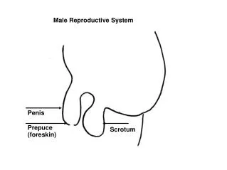
Male Reproductive System. Internal Organs. External organs. Penis scrotum. Testes Various ducts Seminal vesicles Prostate gland Bulbourethral gland. Testes. Paired walnut shaped organs that are located in the scrotum

E N D
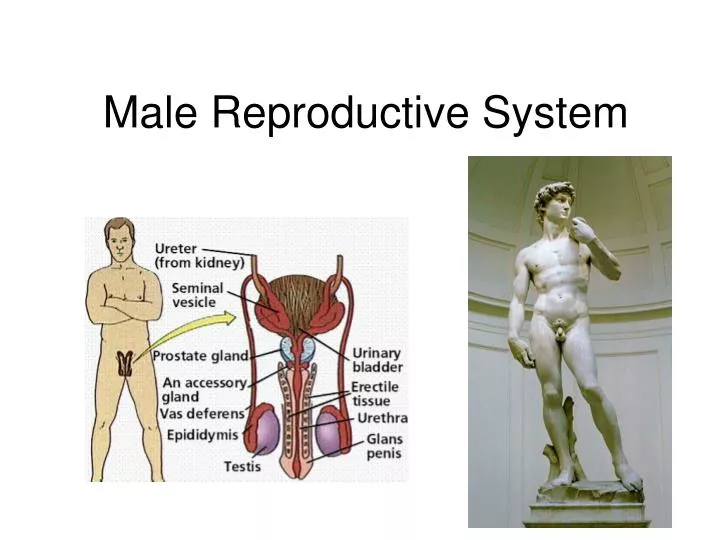
Internal Organs External organs • Penis • scrotum • Testes • Various ducts • Seminal vesicles • Prostate gland • Bulbourethral gland
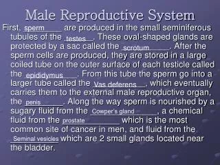
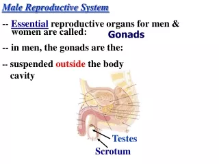
Testes • Paired walnut shaped organs that are located in the scrotum • Consists of highly coiled tubes called seminiferoustubules, where sperm are formed • Leydig cells are scattered between the seminiferous tubules and produce testosterone and other androgens.
Why place the testes outside the body? • Normal sperm development must occur at a lower temperature (2° C lower in scrotum) • Scrotum is a fold of tissue that develops from the abdominal wall cavity • Testes develop high in the abdominal cavity and then descend into the scrotum just prior to birth
In some mammals, such as rodents, the testes are drawn back into the abdominal cavity between breeding seasons. • Some mammals that have low body temperatures (monotremes, whales and elephants) retain their testes within the abdominal cavity permanently
Ducts • From seminiferous tubules, the sperm pass into coiled tubes called the epididymis • It takes about 20 days for sperm to pass through the 6 meter long tubes • As they progress through the epididymis, the sperm become motile (move) and gain the ability to fertilize an egg
During ejaculation, sperm are propelled from the epididymis through the muscular tubes of the vas deferens ducts • The vas deferens run from the scrotum around and behind the urinary bladder, where each joins a duct from the seminal vesicle forming an ejaculatoryduct that opens into the urethra • The urethra runs through the penis to the outside environment (remember that both urine and semen pass through the urethra)
Accessory Glands • Three sets of glands that contribute secretions to form semen (sperm and secretions)
Seminal vesicles- contribute most of the volume of semen. Secretes mucus and fructose, a sugar which provides nutrients to sperm • Prostate gland- secretes anticoagulant enzymes and nutrients • Bulbourethral glands- secretes a mucus that neutralizes any acidic urine left in the urethra
Penis • Composed of 3 cylinders of spongy erectile tissue derived from modified veins and capillaries. • During sexual arousal, the erectile tissue fill with blood from arteries. • As tissue fills, increasing pressure seals off the veins that drain the penis causing it to engorge with blood and stiffen.
Semen • Approximately 2-5 mL of semen is released with each ejaculation and carries 50-130 million sperm. • Once in the female reproductive tract, prostaglandins in the semen cause thinning of the mucus and stimulate uterine contractions to move the sperm toward the waiting egg