Download
1 / 30
340 likes | 476 Views
Development of Female Genital System. Prof. Musaad Al-Fayez. Dr.Jamila EL-Medany. Objectives. By the end of the lecture, you should be able to: Describe the development of gonads ( indifferent & different).

E N D
Development of Female Genital System Prof. Musaad Al-Fayez Dr.Jamila EL-Medany
Objectives • By the end of the lecture, you should be able to: • Describe the development of gonads (indifferent & different). • Describe the development of the female gonad (ovary) and the internal genital organs (uterus, uterine tubes and vagina). • Describe the development of the external genitalia. • List the main congenital anomalies of female genital system.
Development of Genital System • Y chromosome. • Testis-determining factor (TDF) gene. • TDF = Male. • No TDF = Female.
Gonads • Sex of the embryo is determined genetically at the time of fertilization. • Gonads do not acquire male or female morphological characteristics until the 7th week of development.
Gonads • Genital system are developed from two longitudinal ridges of mesoderm which run down the entire length of the dorsal body wall. • These ridges are called urogenital ridges. • The medial region of this ridge differentiates into the genital ridgewhere the gonads develop.
Gonads • The gonads begin to develop during the 5th week in the genital ridge. • First the gonads are undifferentiated and have a cortex and a medulla.
Gonads • In embryos with an XX chromosome complex, the cortex normally differentiates into an ovary, and the medulla regresses. • In embryos with an XY complex, the medulla differentiates into a testis and the cortex regresses.
Gonads • Large primitive cells, called primordial sex cells formed in the yolk sac during the 4th week. • They migrate along the dorsal mesentery of the hindgut to the genital ridges where they become incorporated into the developing gonads.
Gonads • Germ Cells arrives at 5thweek & invades the genital ridge in the 6th week. • During arrival of Germ cells, the epithelium of the genital ridge proliferates, and epithelial cells penetrate the underlying mesenchyme • Forming an irregular shaped cord primitive sex cord “indifferent gonad”
OVARY • Primitive sex cords dissociate into irregular cell cluster. • Later they will disappear and are replaced by vascularstroma forming ovarian medulla. • Surface epithelium of the female gonad proliferates. • In 7th week they give rise to 2nd generation of cords called “cortical cords”.
OVARY • The cortical cords penetrate the underlying mesenchyme. • In 4th month these cords split into isolated cell clusters which surrounding one or more primitive germ cells, (GC). • GC will develop into oogonia and the surrounding epithelial cells form the follicular cells.
GENITAL DUCTS • Two pairs of genital ducts develop in both sexes: 1.Mesonephric (Wolffian) ducts and 2. Paramesonephric (Mullerian) ducts.
GENITAL DUCTS • In female embryos, mesonephric ducts regress while the paramesonephric ducts develop into: • Uterus, • Uterine tubes and • Upper vagina.
Genital duct in the female • Paramesonephric ducts develop into the main genital ducts of the female. • Initially, 3 parts can be recognized in each duct: • A vertical cranial portion (1) which opens into abdominal cavity. • A horizontal part that crosses anterior to the mesonephric duct, (2). 1 1 2 3 2 3
Genital duct in the female • A caudal vertical (3) part that fuses with its partner from the opposite side. • (1 & 2) develop into the uterine tubes. • The fused parts (3) give rise to body and cervix of the uterus. • Mesenchyme will form the myometrium & perimetrium of the uterus.
Vagina • After solid tip of paramesonephric ducts reaches the urogenital sinus, 2 solid evagination grow out. • These evagination (sinovaginal bulbs), proliferate and form vaginal plate. • By the 5th week the outgrowth is entirely canalized.
External genitalia • By the 3rd week, mesenchymal cells migrate around the cloacal membrane to form a pair of cloacal folds. • Cranial to the cloacal membrane the folds unite to form the genital tubercle, (GT). • Caudally the folds are subdivided into urethral folds anteriorly, (UF) and anal folds posteriorly, (AF).
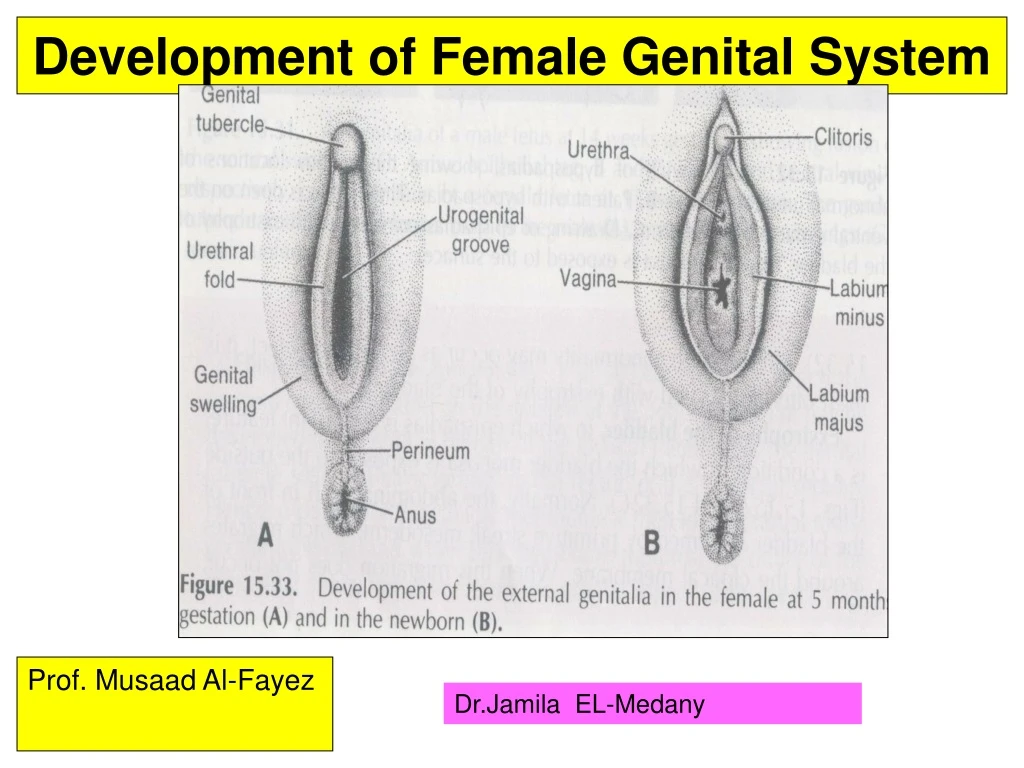
Development of Female External Genitalia • Proliferation of Mesenchyme at the cranial end and sides of the cloacal membrane, will forms: • 1. Genital Tubercle, (GT). • 2. Urogenital Folds or Urethral Folds, (UF). • 3. Labioscrotal swellings or Genital Swellings, (GS).
Feminization of External Genitalia • Estrogenproduced by both placenta and fetal ovaries has a role in feminization of the external genitalia. • Genital Tubercle proliferates to form the primordial Phalls which elongates slightly to form the Clitoris. • Urethral Folds do not fuse and form the Labia Minora. • Labioscrotal Folds form theLabia Majora ,they fuse to form posterior and anterior Labial Commissures.
Congenital Anomalies • Various types of anomalies can result due to: • Arrest of development of the uterovaginal primordium during the 8th week. • Incomplete development of the paramesonephric ducts. • Incomplete fusion of the paramesonephric ducts. • Failure of parts of one or both paramesonephric ducts to develop. • Incomplete canalization.
Cervical Atresia: • It may be combined with incomplete development of the upper vagina or lower uterus. • Vaginal Anomalies: • Atresia(Partial or complete). • Double vagina. • Transversely septate vagina: • Results from faulty canalization of the fused müllerian ducts.
THANK YOU Remnants of mesonephric (wolffian) ducts may persist in the anterolateral wall of vagina or adjacent to the uterus within the broad ligament or mesosalpinx.