Download
1 / 13
150 likes | 390 Views
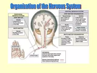
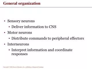
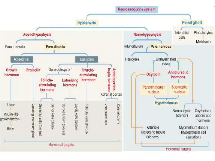
General organization of the neuroendocrine system.

E N D
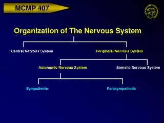
General organization of the neuroendocrine system. The hypothalamus and the hypophysis (pituitary gland) form an integrated system known as the hypothalamohypophysial system consisting of two components: (1) the hypothalamic adenohypophysial system (linking the hypothalamus to the anterior hypophysis), and (2) the hypothalamic neurohypophysial system (connecting the hypothalamus to the neurohypophysis). Functional aspects of the neuroendocrine system. The hypothalamus contains clusters of neurons called nuclei. Some of the neurons are neuroendocrine cells exerting positive and negative effects on the two components of the hypophysis. These effects are mediated by releasing and inhibitory hormones or factors.The transport of signaling molecules is mediated by the hypothalamohypophysial portal circulation consisting of a primary capillary plexus in the lower hypothalamus connected by portal veins to a secondary capillary plexus in the anterior lobe of the hypophysis. A third capillary plexus supplies the neurohypophysis.The primary capillary plexus is supplied by the superior hypophysial artery; the third capillary plexus is supplied by the inferior hypophysial artery. The two arteries are connected by the trabecular artery. There is no connection between the secondary and third capillary plexuses. The hypophysial vein drains the second and third capillary plexuses to the dural sinuses. The hypophysis consists of two embryologically distinct portions: (1) the adenohypophysis or glandular component, derived from Rathke's pouch, an invagination of the roof of the future oral cavity, and (2) the neurohypophysis or neural component, an infundibular downgrowth from the floor of the diencephalon.
The adenohypophysisconsists of three subdivisions: (1) the pars distalis (anterior lobe), (2) the pars tuberalis, surrounding the neural infundibular stem or stalk, and (3) the pars intermedia (the rudimentary intermediate lobe). The neurohypophysis consists of two subdivisions: (1) the pars nervosa and (2) the median eminence.The anterior lobe contains three components: (1) epithelial cell cords, (2) a connective tissue stroma, and (3) fenestrated capillaries (sinusoids) of the secondary capillary plexus.There are three distinct cell populations: (1) acidophil cells (stain with an acidic dye), (2) basophil cells (stain with a basic dye), and (3) chromophobe cells (lacking cytoplasmic staining). Acidophil cells secrete peptide hormones (growth hormone and prolactin); basophils secrete glycoprotein hormones (gonadotropins FSH and LH, TSH, and ACTH). Chromophobe cells are cells that have depleted their cytoplasmic hormonal content. Growth hormone (also called somatotropin). It is secreted in a pulsatile pattern with peak secretion occurring during the first 2 hours of sleep. Growth hormone exerts its actions through insulin-like growth factor-1 (IGF-1) produced in hepatocytes after stimulation by growth hormone. The release of growth hormone is stimulated by growth hormone-releasing hormone produced in the hypothalamus and by high blood levels of IGF-1. Inhibition of growth hormone release is mediated by somatostatin (also produced in the hypothalamus and in the islets of Langerhans in the pancreas) and high blood levels of glucose.Gigantism during childhood and puberty is caused by excessive secretion of growth hormone (usually produced by a benign tumor of the hypophysis called adenoma). Acromegaly (enlargement of hands, feet, jaw, and soft tissues) is seen in adults when growth hormone production is high. Prolactin has a main function: to stimulate the initiation and maintenance of lactation postpartum. Lactation involves (1) mammogenesis, the growth and development of the mammary glands, (2) lactogenesis, the initiation of lactation; and (3) galactopoiesis, the maintenance of milk production. A secondary function is to facilitate the steroidogenic action of LH in Leydig cells by up-regulating the expression of the luteinizing hormone (LH) receptor. The pulsatile secretion of prolactin is regulated primarily by an inhibitory mechanism rather than by stimulation. The main inhibitor is dopamine. Prolactin-releasing hormone and thyrotropin-releasing hormone, both originating in the hypothalamus, stimulate prolactin release.Excessive secretion of prolactin (hyperprolactinemia) by a benign tumor of the hypophysis in both genders causes gonadotropin deficiency. In women, hyperprolactinemia is associated with infertility, anovulation, and oligomenorrhea or amenorrhea (dysfunctional uterine bleeding). A decrease in fertility and libido is seen in males. Galactorrhea (non-puerperal milk secretion) caused by hyperprolactinemia is common in both genders.
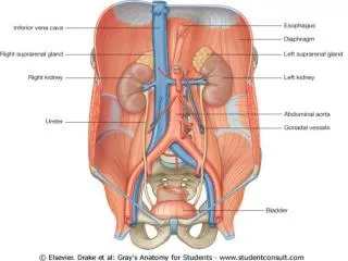
Gonadotropins: FSH and LH. The release of gonadotropins is stimulated by gonadotropin-releasing hormone (GnRH; also called luteinizing hormone-releasing hormone or LHRH). GnRH is secreted in pulses at 60- to 90-minute intervals. A single basophil can produce both FSH and LH.In the female, FSH stimulates folliculogenesis (the development of the ovarian follicle). In the male, FSH targets Sertoli cells in the testes to convert testosterone into estrogen (by aromatization) and produce androgen-binding protein (ABP).In the female, LH stimulates steroidogenesis in the ovarian follicle and corpus luteum. In the male, LH controls the production of testosterone by Leydig cells.The release of FSH and GnRH is inhibited by inhibin (an αβ heterodimer) produced by the target cells (follicular cells and Sertoli cells), and estradiol. The release of FSH is enhanced by activin (a ββ homodimer).A drop in the secretion of GnRH (caused by anorexia nervosa, a tumor of the hypophysis, or a condition known as hypogonadotropic hypogonadism in males) can abolish the secretion of FSH and LH. Castration (ovariectomy or orchidectomy) causes a significant increase in the synthesis of FSH and LH and the vacuolization of gonadotropin-secreting cells (castration cells). Thyroid-stimulating hormone (TSH; or thyrotropin) regulates thyroid function. Thyrotropin-releasing hormone stimulates the release of TSH (and prolactin). Thyroid hormones triiodothyronine (T3) and thyroxine (T4) inhibit the release of TSH.Hypothyroidism, characterized by reduced cell metabolism and temperature, is caused by deficient secretion of TSH and by the autoimmune disorder Hashimoto's disease. Hyperthyroidism is usually determined by an autoantibody directed against the TSH receptor in thyroid follicular cells (Graves' disease). Adrenocorticotropic hormone (ACTH; or corticotropin) stimulates growth and steroid synthesis in the zona fasciculata and zona reticularis of the adrenal cortex.ACTH derives from the large precursor pro-opiomelanocortin (POMC) processed in the anterior hypophysis. Corticotropin-releasing hormone (CRH) derived from neuroendocrine neurons of the paraventricular nuclei (which also produce antidiuretic hormone [ADH]), stimulates the release of ACTH. This CRH stimulatory effect is potentiated by ADH and angiotensin II. High levels of cortisol prevent the release of CRH or ACTH.Cushing's disease, caused by an ACTH-producing adenoma of the hypophysis, results in the overproduction of cortisol by cells of the zona fasciculata of the adrenal cortex, obesity, osteoporosis, and muscle wasting.
Neurohypophysis Three histologic components are found in the neurohypophysis: (1) pituicytes, astrocyte-like cells containing the intermediate filament protein glial fibrillary acidic protein and providing support to axons; (2) unmyelinated axons derived from neuroendocrine cells of the hypothalamic supraoptic and paraventricular nuclei forming the hypothalamic hypophysial tract; and (3) fenestrated capillaries.Axons display intermittent bulging segments called Herring bodies containing neuroendocrine secretory granules. Each secretory granule consists of two components: the carrier protein neurophysin and the associated hormone ADH (also called arginine vasopressin) or oxytocin.Oxytocin participates in the contraction of uterine smooth muscle during labor, and of myoepithelial cells in the lactating mammary alveoli to facilitate milk ejection. ADH regulates water excretion in the kidneys and, at a higher concentration, is also a potent vasoconstrictor.Neurogenic diabetes insipidus occurs when the secretion of ADH is reduced. It is caused by severe head injury, an invasive tumor disrupting the hypothalamic hypophysial tract, or the autoimmune destruction of ADH-producing neurons. Polyuria is a common clinical finding. Nephrogenic diabetes insipidus occurs in certain chronic renal diseases that are not responsive to ADH. Pineal gland. The pineal gland is an endocrine organ containing cells with a neurosecretory function and without direct nerve connection with the brain. The pineal gland is supplied by postganglionic sympathetic nerve fibers derived from the superior cervical ganglia (SCG). Preganglionic fibers to the SCG derive from the lateral column of the spinal cord.
The pineal gland • develops from a saccular outpocketing of the posterior diencephalic roof in the midline of the third ventricle. It contains cells called pinealocytes, arranged in cords and clusters, and supporting glial-like interstitial cells. The pinealocyte displays cytoplasmic extensions with bulbar endings. These cell processes end close to a capillary. Pinealocytes contain abundant mitochondria and characteristic multiple ribbon synapses. Remember that ribbon synapses are also seen in photoreceptor cells of the retina and in hair cells of the inner ear. An important landmark of the pineal gland are calcified deposits called corpora arenacea ("brain sand").The major secretory product of the pineal gland is melatonin, synthesized from tryptophan by pinealocytes and immediately secreted. The concentration of melatonin in the pineal gland is high during the night.The 24-hour circadian clock is an endogenous oscillator controlling circadian rhythms, including sleep and feeding patterns. The retinohypothalamic tract conducts light signals from the retina (in particular from melanopsin-producing ganglion cells that function as luminance detectors) to the hypothalamic suprachiasmatic nucleus (regarded as the circadian "clock"). This is the first regulatory step of melatonin synthesis and secretion.Jet lag, a condition associated with fatigue, insomnia, and disorientation experienced by many travelers, is caused by a disruption of the circadian rhythm. Bipolar disorder and sleep disorder are also linked to the abnormal functioning of the circadian rhythms.A tumor of the pineal gland (called pinealoma) is associated with precocious puberty (pubertas precox) and with a neurologic disorder known as Parinaud's syndrome (paralysis of upward gaze, looking steadily in one direction, pupillary areflexia to light, paralysis of convergence, and wide-based gait).