Download
1 / 10
0 likes | 5 Views
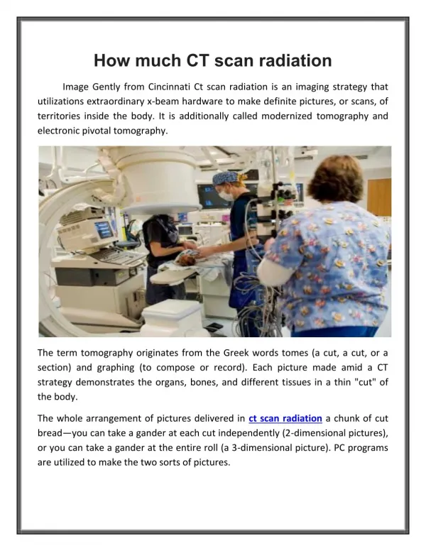
CT scans use ionizing radiation, specifically X-rays, to generate detailed images of the body's internal structures.

E N D
CT Scan Safety: What You Need to Know About Radiation Risks and Precautions
Introduction to CT Scans A CT scan (Computed Tomography scan) is a medical imaging procedure that combines X-rays and computer technology to create detailed images of the body's internal structures. Unlike traditional X-rays, which produce flat, two-dimensional images, CT scans generate cross-sectional (slice-like) images that offer a more comprehensive view of tissues, organs, and bones. These detailed images allow doctors to examine the body in greater depth, helping diagnose a wide range of medical conditions, from fractures and infections to cancers and internal bleeding. CT scans are commonly used in emergency situations due to their ability to quickly assess injuries, and they play a crucial role in pre-surgical planning and monitoring treatment progress for various diseases. The images produced by a CT scan can be used to detect abnormalities in the chest, abdomen, pelvis, and brain, making it an essential tool in modern diagnostic medicine.
How CT Scans Work A CT scan uses a combination of X-ray technology and computer processing to create detailed, cross-sectional images of the body. During the procedure, the patient is positioned on a table that slides into a large, doughnut-shaped machine called a CT scanner. The scanner contains an X-ray tube that rotates around the patient, emitting a series of X-ray beams from different angles. As the X-rays pass through the body, they are absorbed by different tissues to varying degrees. Dense tissues like bones absorb more X-rays, while softer tissues like muscles and organs absorb fewer. A series of detectors inside the scanner measures the amount of X-ray energy that passes through the body, and this information is sent to a computer. The computer then processes this data and constructs a detailed cross-sectional image (or slice) of the body. These slices are stacked together to create a 3D image of the area being scanned. This allows doctors to view and analyze the body's internal structures from multiple angles, helping to identify any abnormalities, such as tumors, fractures, or infections.
Radiation in CT Scans CT scans use ionizing radiation, specifically X-rays, to generate detailed images of the body's internal structures. During the scan, an X-ray tube emits radiation that passes through the body and is detected by sensors, which then create cross-sectional images of tissues and organs. Unlike traditional X-rays, which provide flat images, CT scans offer more detailed, three-dimensional images due to their ability to capture slices of the body from multiple angles. The amount of radiation used in a CT scan is typically higher than in a standard X-ray, as it requires a series of X-ray images to be taken in rapid succession. The radiation dose varies depending on the type of scan and the area of the body being imaged. For instance, a head CT scan uses less radiation than an abdominal scan. While the radiation from a single CT scan is generally considered safe, repeated exposure over time can increase the cumulative radiation dose, which may elevate the risk of developing cancer. Therefore, it’s important for medical professionals to carefully evaluate the necessity of a CT scan, considering factors such as the patient's medical history and potential benefits of the scan.
Risks of Radiation Exposure Radiation exposure during CT scans, while generally safe for most patients, carries some potential risks, especially when the exposure is repeated over time. The primary concern with radiation from CT scans is its potential to damage cells or DNA, which can increase the risk of developing cancer later in life. However, the risk from a single CT scan is typically small, but it increases with the number of scans a person undergoes, particularly over long periods. Short-Term Risks: • For the most part, the radiation dose in a CT scan is brief and localized, meaning it does not cause immediate harm or noticeable side effects. Short-term risks are minimal, but caution is advised with repeated or unnecessary scans. Long-Term Risks: • The long-term risks of radiation exposure are more concerning. Repeated exposure to ionizing radiation can lead to accumulated doses over time, which may increase the likelihood of developing cancer. For instance, studies have shown that excessive radiation exposure over time can slightly raise the risk of leukemia or solid cancers, such as those of the brain, lung, or thyroid.
Minimizing Radiation Exposure Minimizing radiation exposure during CT scans is crucial to reducing potential health risks while ensuring the procedure remains effective for diagnosis and treatment. Several strategies are employed to limit radiation dose without compromising the quality of the images. 1. Low-Dose CT Scanning: • Low-dose technology allows for the use of reduced radiation levels while still maintaining the necessary image quality for accurate diagnosis. Newer CT machines are designed to deliver lower doses of radiation, especially for routine scans, without sacrificing detail. 2. Adjusting Scan Parameters: • Scan settings can be adjusted to ensure the most appropriate radiation dose is used. Factors such as beam strength, scan duration, and number of slices can be tailored to the patient’s size, the area being scanned, and the purpose of the scan. For example, pediatric CT scans often use lower doses due to children’s increased sensitivity to radiation.
Safety Precautions for Patients To ensure the safety and well-being of patients during a CT scan, several precautions are taken to minimize radiation exposure and prevent any potential complications. These measures help ensure that the scan is both necessary and as safe as possible. 1. Informing Healthcare Providers: • Provide Medical History: Patients should inform their healthcare provider about any prior CT scans, allergies, or existing medical conditions. This helps doctors assess the necessity of the scan and choose the safest approach. • Disclose Pregnancy: It is especially important for women to inform the healthcare team if they are pregnant or suspect they may be. While CT scans are generally avoided during pregnancy, alternative imaging methods like ultrasound or MRI are often preferred to avoid potential risks to the fetus. 2. Discussing the Need for the Scan: • Evaluate Necessity: Healthcare providers should evaluate whether a CT scan is the best option for diagnosing the condition or if another imaging method (such as an MRI or ultrasound) might be safer or equally effective. • Use of Contrast Agents: If a contrast dye is required for the scan, patients should inform their doctor about any known allergies or kidney problems, as contrast agents can have side effects in certain individuals.
The Role of Medical Professionals Medical professionals play a crucial role in ensuring that CT scans are used effectively, safely, and appropriately. From determining the need for a scan to interpreting the results, each step involves careful decision-making to balance diagnostic accuracy with patient safety. 1. Evaluating the Need for a CT Scan: • Doctor’s Assessment: The physician (often a general practitioner or specialist) is responsible for evaluating whether a CT scan is necessary. They assess the patient’s symptoms, medical history, and potential benefits of the scan. In some cases, alternative imaging methods, such as MRI or ultrasound, may be preferred to avoid radiation exposure. • Risk vs. Benefit Analysis: Medical professionals carefully consider the risks of radiation exposure and weigh them against the potential diagnostic benefits. This is particularly important in vulnerable populations, such as pregnant women and children. 2. Conducting the Scan: • Radiologic Technologists: These trained professionals are responsible for operating the CT scanner. They position the patient correctly, ensure the machine is calibrated, and follow protocols to obtain clear and accurate images while minimizing radiation exposure. They also monitor the patient during the scan to ensure comfort and safety.
Advances in CT Technology Recent advancements in CT technology have significantly improved the quality of imaging, reduced radiation exposure, and enhanced the overall efficiency of the procedure. These innovations are transforming how medical professionals use CT scans for diagnosing and monitoring diseases, allowing for more precise and safer patient care. 1. Low-Dose CT Scanning: • Minimized Radiation Exposure: One of the most significant advances in CT technology is the development of low-dose CT systems, which provide high-quality images while using significantly less radiation. These systems are particularly useful for sensitive patient groups, such as children and individuals who need multiple scans over time. New algorithms and software help optimize the scanning process, reducing radiation doses without sacrificing diagnostic accuracy. 2. Faster Scanning Speed: • Improved Imaging Efficiency: Advances in scanner design and computer processing power have dramatically reduced the time it takes to conduct a CT scan. Modern CT scanners can capture detailed images in a fraction of the time compared to older models, which is especially beneficial for emergency situations where rapid diagnosis is essential. Faster scanning also helps improve patient comfort and reduces motion artifacts from breathing or movement during the scan.
Conclusion and Key Takeaways CT scans are essential tools in modern medicine, providing fast, accurate, and detailed images that help diagnose a wide variety of medical conditions. While they offer significant benefits in terms of diagnostic capabilities, it’s important to recognize the associated risks, particularly the exposure to ionizing radiation. By understanding these risks and following safety guidelines, the benefits of CT scans can be maximized while minimizing potential harm. Key Takeaways: • CT Scans Are Crucial for Diagnosis: CT scans are invaluable for diagnosing conditions ranging from fractures and infections to tumors and internal bleeding. Their ability to provide detailed, cross-sectional images makes them essential in emergency care and complex medical cases. • Radiation Exposure Is a Consideration: While the radiation used in CT scans is necessary for producing clear images, it does carry a risk. Although the exposure from a single scan is generally considered low, the cumulative effects of repeated scans over time can increase the risk of cancer. • Minimizing Radiation: Advancements in technology, such as low-dose CT scans, improved imaging techniques, and AI integration, help reduce radiation exposure without compromising image quality. Medical professionals also adjust scan parameters to ensure the lowest possible dose for the patient.