Download
1 / 49
611 likes | 1k Views
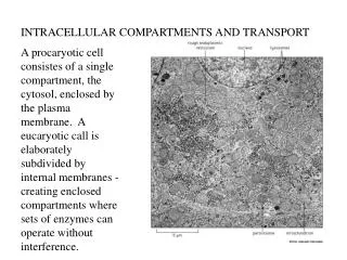
Intracellular Compartments and Transport. ECB Ch 15. In eucaryotic cells, internal membranes create enclosed compartments and organells in which different metabolic processes are segregated. 15_01_organelles.EM.jpg. part of a liver cell. glycosomes.

E N D
Intracellular Compartments and Transport ECB Ch 15
In eucaryotic cells, internal membranes create enclosed compartments and organells in which different metabolic processes are segregated. 15_01_organelles.EM.jpg part of a liver cell glycosomes
basic set of organells found in most animal cells 15_02_cell_intestine.jpg endomembrane system (double membrane, nuclear envelope) nuclear pore
(new membrane synthesis) P498, 499 cytoplasm?
15_03_ER_evolved.jpg prokaryotes: small size, high surface-to-volume ratio plasma membrane provide all membrane-dependent functions
15_04_Mito_origin.jpg (mitochondria and chloroplasts possess their own small genomes and can make some of their own proteins)
protein sorting 15_05_import_proteins.jpg by protein translocators (protein unfolded)
Signal sequence: a continuous stretch of a. a. sequence,15-60 a.a. long; necessary and sufficient to direct a protein to a particular organell.
Nuclear envelope 15_07_out_nucl_memb.jpg nuclear lamina: a fine woven meshwork of protein filaments that lines the inner face of inner nuclear membrane and provides a structural support for the nuclear envelope • The inner nuclear membrane contains proteins that act as binding site for the chromosomes and for the nuclear lamina. • The outer nuclear membrane is continuous with the ER.
15_08_nuclear_pore.jpg • nuclear pore: composed of ~100 proteins • water-soluble molecules- nonselective • RNAs, proteins and macromolecular complexes- selective (sorting signals)
(having the appropriate sorting signals: nuclear location signals) (Table 15-3) 15_09_pore_transport.jpg (fully folded) (guide the complex into the pore) active transport: energy provided by GTP hydrolysis (open the nuclear pore just the right amount to allow the protein complex to pass through) transport receptor -mRNA
Transportation of proteins across organelles membranes • nucleus: fully folded conformation proteins, ribosomal compartments as assembled particles • other organelles: • mitochondria, chloroplasts, ER, …: unfolded protein • peroxisomes: unclear
An unfolded protein is imported into a mitochondrion 15_10_unfolded_imprt.jpg • usually have their signal sequence at the N-terminus (by a signal peptidase) (the chaperone proteins that help to pull the protein across the membrane and help it to refold)
The mitochondria and chloroplasts membranes: • The growth and maintenance of mitochondria and chloroplasts requires not only the import of new proteins but also the incorporation of new lipids into their membranes. • Most of their membrane phospholipids are thought to be imported from the ER (the main site of lipid synthesis in the cell). • Phospholipids are transported individually to the mitochondria and chloroplasts by water-soluble lipid-carrying proteins. • These proteins ensure that the different cellular membranes retain their characteristic lipid composition.
ER: the most extensive membrane system in a eucaryotic cell • served as an entry point for proteins destined for other organelles (Golgi apparatus, endosomes, lysosomes), for cell surface (plasma membrane) and cell exterior, as well as for the ER itself. 15_11_ER.jpg • Two kinds of proteins are transferred from the cytosol to the ER: • water-soluble proteins: completely into the ER lumen • transmembrane proteins:
Two kinds of proteins are transferred from the cytosol to the ER: • water-soluble proteins (completely translocated across the ER membrane and released into the ER lumen) • for secretion, or for the lumen of an organelle • transmembrane proteins (partly translocated across the ER membrane and become embedded in it) • to resides in the ER membrane, the membrane of another organelles, or the plasma membrane • all of these protein are initially directed to the ER by an ER signal sequence, a segment of 8 or more hydrophobic a.a. • also involved in the process of translocation across the membrane
15_12_pool_ribosomes.jpg free ribosomes (identical) RER Most of the proteins that enter the ER begin to be threaded across the membrane before the polypeptide chain is completely membrane-bound ribosomes rough ER
The ER sequence is guided to the ER membrane with the aid of at least tow components: • a signal-recognition particle (SRP), present in the cytosol, which binds to the ER signal sequence when it exposed on the ribosome • an SRP receptor, embedded in the membrane of the ER, which recognizes the SRP
The ER sequence is guided to the ER membrane with the aid of at least tow components: 15_13_ER_signal_SRP.jpg ( binding of SRP slow down the protein synthesis , until they further bind to an SRP receptor that the synthesis recommences) SRP & SRP receptor function as molecular matchmakers, connecting ribosomes that are synthesizing proteins containing ER signal sequences to available ER translocation channels.
Water-soluble protein: 15_14_enters_lumen.jpg • The signal sequence of Water-soluble protein: • almost always at the N-terminus • functions to open the translocation channel
Transmembrane protein 15_15_into_ER_membr.jpg with a defined orientation Start and stop signals determine the arrangement of a transmembrane protein in the lipid bilayer
Double-pass transmembrane protein 15_16_double_pass.jpg • Start transfer sequence • internal signal sequence • never removed from the polypeptide
Vesicular transport 15_17_Vesicles_bud.jpg (endocytic pathway) (secretory pathway) All of the recognition events depend on proteins associated with the transport vesicle membrane.
Vesicle budding is driven by the assembly of a protein coat 15_18_Clathrin_EM.jpg clathrin-coated pit dynamin (a small GTP-binding protein) coated vesicle: • clathrin-coated vesicles: • the well -studied coated cesicles • bud from Golgi apparatus on the secretory pathway and from the plasma membrane on the endocytic pathway • function of the coat: • shape the membrane into a bud • helps to capture molecules for onward transport Clathrin coated pits and vesicles budding from the plasma membrane of cultured skin cells
15_19_Clathrin_vesicle.jpg (with specific transport signals) There are at least two types of adaptins: the adaptins that bind cargo receptor in the plasma membrane are different from those that bind cargo receptors in the Golgi apparatus.
COP-coated vesicles (COP: coat protein): a different class of coated vesicles involved in transporting molecules between the ER and Golgi apparatus and from one part of the Golgi apparatus to another.
SNAREs (a family of related transmembrane proteins) help direct transport vesicles to their target membranes 15_20_SNAREs.jpg (a specific marker protein) v: vesicle t: target according its origin and cargo
SNARE proteins play a central role in membrane fusion 15_21_membr_fusion.jpg • after docking, it often awaits a specific molecular signal then fusion • all membrane fusions in cells are catalized by specific proteins that assemble at the fusion site to form a fusion complex that provides the means to cross the energy barrier for fusion process Pairing of v-SNAREs and t-SNAREs forces the two lipid bilayers into close apposition (< 1.5 nm). Lipids then flow between the two bilayers and the membrane fuse.
Most proteins are covalently modified in the ER • disulfide bonds formation • formed by the oxidation of pairs of cysteine side chain catalyzed by an enzyme that resides in the ER luman • glycosylation • formed by the covalent attachment of short oligosaccharide side chains carried out by glycosylating enzymes found in the ER • this oligosaccharide processing begins in the ER and continues in the Golgi apparatus
Many proteins are glycosylated in the ER 15_22_glycosylated_ER.jpg asparagine-X-serine asparagine-X-threonine • N-linked glycosylation: • oligosaccharide side chains linked to an Asn NH2 group in a protein • the most common type of linkage of found on glycoproteins (transfer as an intact unit) oligosaccharide processing
Exit from the ER is controlled to ensure protein quality 15_23_Chaperones.jpg Chaperones prevent misfolded or partially assembled proteins from leaving the ER. misfolded protein: refolding or degradation ER retention signal
Proteins are further modified and sorted in the Golgi apparatus. ER (entry) 15_24_Golgi .jpg (exit) plasma membrane
the regulatory and constitutive pathway of exocytosis in secretory cells 15_28_trans_Golgi_net.jpg acidic pH and high Ca2+
constitutive exocytosis pathway (default pathway) • operates continually in all eucaryotic cells • proteins not are aggregated • supplies newly made lipids and proteins to the plasma membrane for its growth • after secretion • becoming peripheral proteins • incorporated into the extracellular matrix • diffusing into the extracellular fluid • signaling other cells
regulated exocytosis pathway • operates only in secretory cells • secretory cells produce hormones, mucus, digestive enzymes, which are sorted in secretory vesicles (sorted packaged in the trans Golgi network) for release • proteins in secretory vesicles are aggregated to form high concentration
15_29_Secretory_vesicl.jpg discharge concentrated aggregates of protein
endocytosis • pinocytosis (cellular drink) • ingestion of fluid and molecules via small vesicles (d<150 nm) (continually in all eucaryotic cells) • phagocytosis (cellular eating) • ingestion of large particles (microorganisms, cell debris) via large vesicles (phagosomes) (d>250 nm) (mainly by specialized phagocytic cells) • pseudopods
15_30_white_bloodcell.jpg form a phagosome and then fuse with a lysosome and the microbe is digested
A macrophage 15_31_macrophage.jpg pseudopods
Cell membrane is added by exocytosis and removed by endocytosis • Pinocytosis is mainly carried out by clathrin-coated pits and vesicles • Vesicle fuse with an endosome
Receptor-mediated endocytosis 15_32_LDL_enters.jpg Some others transported by receptor-mediated endocytosis : Vit B12, iron, extracellular signaling molecules, viruses LDL: low-density lipoproteins
The fate of receptor proteins involved in endocytosis depends on the type of receptor. • early endosomes: • just beneath the plasma membrane • late endosomes: • 5-15 min later, fuse with one another or a preexisting late endosome 15_33_type_of_receptor.jpg The enterior of the endosome compartment is kept acidic (pH 5-6) by an ATP-driven H+ pump p527
lysosome 15_34_lysosome.jpg • the specilized digestive enzymes and membrane proteins of the lysosome are synthesized in the ER and transported through the Golgi apparatus • a mannose 6-phosphate tagged on the enzymes • specifically transport to lysomosome by being recognized of the mannose 6-phosphate receptor ~40 types of hydrolytic enzyme most of the lysosomal membrane proteins are highly glycosylated, to protected them from digestion by the lysosomal proteases
Materials degradation 15_35_path_lysosome.jpg