Download
1 / 52
600 likes | 1.58k Views
Lung tissue Pneumonia Pulmonary hemorrhage Pulmonary edema Respiratory distress syndrome (hyaline membrane disease). Causes of Respiratory Failure I. wet lung. HMD. meconial aspiration. congenital pneumonia. Adults and children: Acute respiratory distress syndrome (ARDS).

E N D
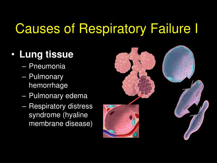
Lung tissue Pneumonia Pulmonary hemorrhage Pulmonary edema Respiratory distress syndrome (hyaline membrane disease) Causes of Respiratory Failure I
wet lung HMD meconial aspiration congenital pneumonia
Adults and children: Acute respiratory distress syndrome (ARDS) Mortality: 25 - 35% Newborn: Infant respiratory distress syndrome (iRDS) CLD: 15 - 25% Oxygenation Lung volumes Pulm. compliance Mechanical ventilation Ventilator induced lung injury
MRI signal intensity from non-dependent to dependent regions The water burden of the lung makes the lung of the preterm infant, despite surfactant treatment,vulnerable to VILI 2-day-old, 38-week gestation infant 4-day-old, 26-week gestation infant Adams EW AJRCCM 2002; 166:397–402
Nonhomogeneous Lung Disease The pathophysiology shared by these diseases is nonuniform lung involvement where certain lung units are nearly normal while other areas are markedly abnormal. A strategy that is effective in opening damaged areas may result in overinflation and trauma to more normal areas of the lung.
Diffuse “Homogeneous” Lung Disease The goals of assisted ventilation in this group of patients are to improve lung inflation, compliance and ventilation/perfusion matching while avoiding barotrauma or compromise of cardiac output.
The best approach = The extended sigh (stepwise increase and decrease of PEEP using the lowest VT possible) Required Monitoring: SaO2, PaO2 PaCO2 and/or endtidal CO2 Hemodynamics
Recruitment Overdistension tidal volume "static" compliance: static PIP (Pplat) - PEEP Cst = PEEP titration The oxygenation response: Can it be used? Burns D J Trauma 2001;51:1177-81
20/5 ETT disconnection PEEP 20 PEEP 15 PEEP 10 PEEP titration: O2 and CO2 response in a lung injury model of surfactant depletion Steps of 5 cmH2O to 35/20 Pressure control ventilation 20/5 PEEP 5
b) flow diversion VA VA PaO2 PaO2 PaCO2 PaCO2 O2-improvement = Shunt improvement = a) recruitment
Prevalent overinflation = dead space effect 1 1 2 2 1 1 1 1 1 1 – – PEEP 5 PEEP 15 PaO2 and PaCO2 increase O2-improvement does not exclude overinflation Gattinoni L (2003)
Allowable Vt and disease severity ALI (surfactant depleted lung) Volume (l) severe (A)RDS Airway pressure (cmH2O)
Transition from CMV to HFOV • Pplat approaching 25 cmH2O after PEEP trial (recruitment) • and / or PEEP > 12 cmH2O 2) Reduction of Vt < 5 required to match Pplat limits 3) “uncomfortably” high pCO2 or low pH (level dependent from additional pathologies)
CMV CMV HFOV HFOV Rationale for HFOV-based lung protective strategies HFOV uses very small VTs. This allows the use of higher EELVs to achieve greater levels of lung recruitment while avoiding injury from excessive EILV. 2.Respiratory rates with HFOV are much higher than with CV. This allows the maintenance of normal or near-normal PaCO2 levels, even with very small Vts.
The concept of volume recruitment during HFO Suzuki H Acta Pediatr Japan 1992; 34:494-500
12 11 10 9 11 Continuous blood gas monitoring during HFO CDP: 13 Overdistention Collapse
Lung hypoplasia syndromes Congenital diaphragmatic hernia Potter syndrome prolonged rupture of membranes Hydrops fetalis Causes of Respiratory Failure II • The common variable in this group of infants is small, often abnormal lungs. This is associated to: • Difficult CO2 elimination • Pulmonary hypertension (PPHN)
Congenital diaphragmatic hernia Gentle ventilation (peak pressure limitation) “Permissive” hypercapnia resp acidosis May worsen PPHN iNO HFO ECMO “Versus” VILI (baro- volutraumatisme)
Congenital diaphragmatic hernia Accept ductal shunting as long as RV function is not impaired! Bohn D Am J Respir Crit Care Med 2002; 166: 911–915
Survival rates in CDH Total Survivors ECMO Bohn D Am J Respir Crit Care Med 2002; 166: 911–915
“Geneva” attitude Surfactant (-) NO +/- (Cardiac US!) HFOV +++ (early) ECMO (-) The Scandinavian Experience with CDH Sakri H Pediatr Surg Int (2004) 20: 309–313
Conducting airways Aspiration (before or after birth) Congenital malformation Tracheal fistula Causes of Respiratory Failure III
+ + Extra- and intrathoracic airway obstruction Stridor From Pérez Fontán JJ, 1990
No PEEP PEEP 10cmH2O Classical pathological conditions that may lead to a difficult to ventilate situation Severe airway compression / malacia courtesy from Quen Mok, Great Ormond Street Hospital for Children, London
Severe airway compression Once you can ventilate these patients (with high PEEP) they are usually difficult to extubate My advice: Keep a high PEEP on spontaneous ventilation, reduce pressure support and extubate from a high PEEP (ev. to CPAP or NIV)
External PEEP in obstructive lung disease (PEEP-trial) Caramez MP Crit Care Med 2005; 33:1519 –1528
External PEEP in obstructive lung disease (PEEP-trial) Caramez MP Crit Care Med 2005; 33:1519 –1528
HFOV in severe airway obstruction Duval E Pediatric Pulmonology 2000: 30:350–353
Air leak syndromes Pneumothorax Bronchopulmonary fistula PIE Causes of Respiratory Failure IV
CMV HFOV Tracheal pressure (cmH2O) Endinspiration Endexpiration CMV HFOV PIP Classical indication for HFV - because of small pressure swings PEEP
PIE, bronchopleural fistula, pneumothroax Recruit to improve oxygenation and in order to lower the FiO2 needed – then reduce the airway pressures to the lowest level needed (air leak will often cease) • References: Shen Chest 2002;121;284-6 • Mayes Chest. 1991; 100:263-4 • Rubio Intensive Care Med. 1986;12:161-3 One sided intubation or airway blocking by inserted balloon catheters is almost never required even in severe airleak (this was just a nice idea to get a case report)
Pulmonary perfusion Congenital heart disease Persistent fetal circulation Causes of Respiratory Failure V
31 6/7 wks GA, 1000 g GA (small for GA) 1 course of prenatal steroids 12 hours before delivery Presents with respiratory distress at birth: RR 64, indrawing, SO2 84% at RA CPAP trial with fast increasing O2 requirements (> 60%) Venous and arterial umbilical catheter First art BGA: pH 7.09, PCO2 11 kPa (83 mmHg), pO2 4.36 Intubation Vent settings: TCPL, RR 60, PEEP 5, PIP 18 Poor sats persists: SO2 78% under FiO2 80%
PIP 24, PEEP 8, RR 60 no real change in SO2 (SaO2 82 % , FiO2 100%) Art BGA: pH 7.11, pCO2 10 kPa, pO2 3.33, BE –3.6 A: Surfactant? B: HFOV? C: Other? Switch to HFOV: CDP 19, Pressure Ampl 46, Freq 12 Hz SO2 80 %, FiO2 100% Art BAG: pH 7.31, pCO2 6.1, pO2 3.56, BE –2.8
A: Surfactant? B: Increase CDP? C: Other? CDP 19, Pressure Ampl 46, Freq 12 Hz SO2 80 %, FiO2 100% Art BAG: pH 7.31, pCO2 6.1, pO2 3.56, BE –2.8
CDP 19, Pressure Ampl 46, Freq 12 SO2 80 %, FiO2 100% Art BGA: pH 7.31, pCO2 6.1, pO2 3.56, BE –2.8 CDP 14, Pressure Ampl 34, Freq 15 SO2 92 %, FiO2 can be lowered fast to 40% Art BGA: pH 7.37, pCO2 5.3, pO2 3.58, BE –1.6 Diagnosis and what next?
CDP 14, Pressure Ampl 34, Freq 15 SO2 92 %, FiO2 40% Art BGA: pH 7.37, pCO2 5.3, pO2 3.58, BE –1.6
CDP 14, Pressure Ampl 34, Freq 15 Hz SO2 92 %, FiO2 can be lowered fast to 40% Art BGA: pH 7.37, pCO2 5.3, pO2 3.58, BE –1.6 SO2 78 % CDP 13, Pressure Ampl 30, Freq 15 Hz SO2 74 % SO2 91 %, FiO2 can be furter lowered to 25% Art BGA: pH 7.42, pCO2 4.4, pO2 3.50, BE –2 iNO 8 ppm CDP 13, Pressure Ampl 25, Freq 15 Hz Echo cardiac SO2 94 %, FiO2 can be furter lowered to 21% Art BGA: pH 7.39, pCO2 4.87, pO2 3.59, BE –2.3
6 hours later (after refixation of ETT) rapid drop in saturation to values around 60 to 65% under FiO2 of 100%, hemodynamic stable (BP 49 / 30) • Increase in airway pressures for recruitment? • Surfactant • Increase iNO concentration • Other BGA:
CDP 13, Pressure Ampl 25, Freq 15 Hz, FiO2 100%, iNO 12 ppm Lactate: 2.2 4.5 Stepwise increase in CDP up to 20 SO2 72% pre and postductal Art BGA: pH 7.22, pO2 3.56, pCO2 8.0, BE - 3 Gradually increase in P-Ampl to 46 Surfactant SO2 varies around 65 to 75% on FiO2 100%, iNO 12 ppm Art BGA: pH 7.1, pCO2 5.0, pO2 2.36, BE - 5
CDP 20, Pressure Ampl 48, Freq 10 Hz, FiO2 100%, iNO 12 ppm SO2 varies around 55 to 75% Art BGA: pH 6.97, pCO2 10.0, pO2 2.86, BE – 12, Lactate 8.6 • Increase iNO, B) switch to CMV • C) change HFO settings, D) second dose of surfactant
CDP reduction from 20 to 14 Sat immediately improves to 90%, allowing to reduce FiO2 to 60 then 40 % Anticipate! A) I have to reduce iNO B) I lower further CDP C) I change other settings – which one? D) Excellent work, I need a coffee now! Reduce pressure amplitude immediately when lowering CDP (coming of overdistension will render oscillation swings more effective!) Pressure amplitude from 48 to 30 (visible wiggeling) Art BGA: pH 7.39, pCO2 3.4, pO2 6.26, BE – 10 CDP reduction from 14 to 10, P-amplitude to 24, FiO2 to 21%
R-L shunt across the FO • severe hypoxemia • RV dilatation and failure • poor CO • Moderate mainly • postductal hypoxemia • + ev R-L shunt FO 2) In general good CO NO yes NO may lead to L-R shunt with pulmonary flooding PPHN with: Open ductus Closed ductus
RDS and PPHN in the newborn infant: Nitric oxide effect Right to left shunt without iNO Left to right shunt on iNO PA PA Duct Duct Ao Ao Indication: not poor postductal oxgygenation but signs of poor cardiac output
Take home messages It is not always iRDS that causes hypoxemia in the preterm infant If you don’t know what to do next with your ventilator settings reduce your airway pressures first Try to anticipate changes in respiratory mechanics and gas exchange before turning knobs on your ventilator
Pressure – Flow – Time - Volume Time constant: T = Crs x Rrs To short Ti and/or Te will lead to inefficient alveolar ventilation and risk of intrinsic PEEP Adapt your respirator rate (Ti and/or Te) to the stage and mechanical characteristics of lung disease The saying “ we ventilate at 60/min” is a testimony of no understanding