Download
1 / 54
550 likes | 724 Views
Explore the anterior aspect of facial bones, including palatine, zygomatic, lacrimal, nasal, vomer, and mandible. Learn about fractures, foreign bodies, osteomyelitis, neoplasms, and TMJ syndrome. Radiography positioning techniques discussed.

E N D
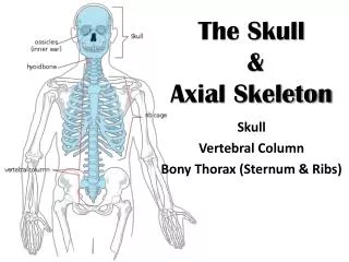
Facial Bone Anatomy & Positioning RTEC 233 Week 6 FINAL
Anterior Aspect of Facial Bones • 6 paired facial bones 1) 2) 3) 4) 5) 6) • 2 single bones 7) 8) 4 3 2 1 7 7 5 8
Palatine Bones • L-shaped bones • _________ portion forms posterior hard palate • _______ portion extends between 1maxillae and 1 pterygoid plate of sphenoid bone • Articulates with 2 cranial bones and 4 facial bones
Zygomatic Bones • Forms ________ • Forms lower outer margin of _______ • Articulates with 3 cranial bones 1) 2) 3) • Articulates with maxillae
Inferior Nasal Cochae • The only pair of conchae that are _______ facial bones • Articulates with 1) 2) 3) 4) • Covered with mucous membranes to: ______________________________________________
Lacrimal Bones • About the size & shape of a fingernail • Lacrimal foramen for tear duct • Lie anteriorly on the medial side of orbit • Can be seen on PA and lateral projections • Articulates with 2 cranial bones and 2 facial bones
Nasal Bones • Fused and form bridge of nose • Vary in size considerably • The point of junction with the frontal bone is the nasion • Articulates with 2 cranial and 2 facial bones
Vomer • Forms inferosuperior part of nasal septum • Deviated nasal septum • Depressions for blood vessels • Articulates with 2 cranial bones & 4 facial bones
Mandible • Only _______ bone in the skull • ________& largest facial bone • ___bones at birth • Contains ______foramina
Fractures Blowout Tripod LeFort Coutrecoup Foreign Body Osteomyelitis Neoplasms Secondary Osteomyelitis TMJ Syndrome Pathologic Indications for Facial Radiography
Positioning: Lateral Facial bones • CR is perpendicular • enters lateral zygomatic bone ½ way between outer canthus and EAM.
Lateral Facial Radiograph • All facial bones in with zygomatic bone in center • Almost SI mandibular rami • SI orbital roofs (no tilt) • No rotation of sella turcica
Positioning: Waters • CR perpendicular to exit acanthion
Waters Radiograph • Distance from lateral border of skull and orbit equal on each side • Petrous ridges projected immediately below maxillary sinuses
Reverse Waters • CR perpendicular and enters acanthion
Reverse Waters Radiograph • Distance from lateral border of skull and orbit equal on each side • Petrous ridges projected immediately below maxillary sinuses
Modified Waters • CR perpendicular and exits acanthion
Modified Waters Radiograph • Petrous ridges projected immediately below the inferior border of the orbits • Equal distance from lateral orbit to lateral skull on both sides
PA Axial - Caldwell • CR 15 caudal to exit nasion
PA Axial- Caldwell Radiograph • Equal distance from lat skull to lat orbit • Symmetric petrous ridges in lower 1/3 orbit • Penetration of frontal bone without excessive density at lateral borders of skull.
Lateral Nasal Bones • CR perpendicular to the bridge of nose at a point 1” distal to the nasion
Lateral Nasal bones Radiograph • No rotation of nasal bone and soft tissue • Anterior nasal spine and frontonasal suture evident • Close collimation