Download
1 / 16
170 likes | 582 Views
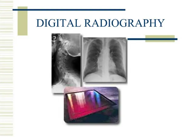
DIGITAL RADIOGRAPHY. Digital Fluoroscopy. Input phosphor output phosphor electronic signal beam splitter video signal TV monitor Video signal is a voltage signal which varies continuously ADC (Analog Digital Converter) converts analog to digital.

E N D
Digital Fluoroscopy • Input phosphoroutput phosphor electronic signal beam splitter video signal TV monitor • Video signal is a voltage signal which varies continuously • ADC (Analog Digital Converter) converts analog to digital
What are some negatives of conventional (film) radiography? • Difficult to image both soft tissue and bony structures in same image • Difficult to differentiate between the subtle differences of soft tissues (shades of gray---long contrast, low contrast) • Unable to gather quantitative info about attenuation characteristics of anatomy • Image is processed permanent as is • Amount of time needed to process • Archiving/storage/ acquisition issues
Similarities between CR and film radiography • Same x-ray tube and generator • Still select optimum kVp and mAs • Accurate positioning • Use cassette or image receptor • There is still a latent image which can be processed into a manifest image
DIFFERENCES • Imaging plate rather than intensifying screen/film • Photostimulable phosphor-europium activated barium fluorohalide phosphor • 200 screen speed equivalent • Phosphors absorb photons • Capable of wider latitudes = better visualization of soft tissues and bone
DIFFERENCES CONT. • Film made of minute strands of black metallic silver • Digital image = rows and columns called a matrix
MATRIX, PIXEL AND VOXEL • Matrix made up of pixels ( picture element) • Pixels = x-ray intensity at that location and given a numeric value for the shade of gray • Voxel represent the volume of tissue of the patient • Matrix preferred size – 2048 x2048 or 4, 194,304 pixels • Larger matrix = more pixels and pixels are smaller
IMAGE ACQUISITION • Exit(remnant radiation) IP photons absorbed photoelectrically by phosphor LATENT IMAGE • Exposed IP reader unit (digitizer) scanned to release energy as light photomultiplier tube (PMT) collects, amplifies and converts light to electronic signal to ADC manifest image
Manifest image is a matrix composed of pixels with assigned brightness levels • IP scanned again with intense light to erase plate • 10,000 readings
Histograms/window or index levels/algorithms • Histogram-graphic display of digital data • Used to evaluate adequacy of IP to x-rays • PMT needs to be adjusted to compensate for errors • Window/index levels – • Algorithms – math formulas needed to formulate image construction based on anatomy imaged • Radiographer must indicate correct procedure so the correct algorithms are used.
IR/IP • List some characteristics of an IP • What speed film screen system is associated with a typical IP? • How does this affect technique?
DR • DIRECT READOUT DIGITAL RADIOGRAPHY • FLAT PANEL DIRECT CAPTURE DETECTOR • CHARACTERISTICS? • COMPARE INDIRECT CONVERSION TO DIRECT
Post processing • Subtraction • Contrast enhancement • Edge enhancement • Black and white reversal • Compensate for errors
Misc. • Resolution =2.5 line pairs per millimeter • Window level = adjusts image brightness • Window width = adjusts radiographic contrast • Quantum mottle • Artifacts • Scatter
PACS • HIS • RIS