Download
1 / 49
490 likes | 700 Views
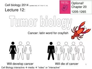
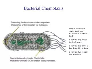
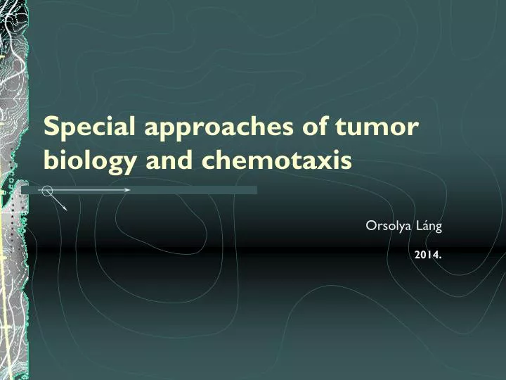
Special approaches of tumor biology and chemotaxis. Orsolya Láng 2014. TUMOR CELLS AND MIGRATION. PRIMARY TUMOR. METASTASIS. Angiogenesis Adhesion. CELL and CELL CYCLE. Oncogenes. Apoptotic molecules. Growth factors. Chemokines. Cell cyle regulatory proteins. Adhesion molecules.

E N D
Special approaches of tumor biology and chemotaxis OrsolyaLáng 2014.
TUMOR CELLS AND MIGRATION PRIMARY TUMOR METASTASIS Angiogenesis Adhesion
Oncogenes Apoptotic molecules Growth factors Chemokines Cell cyle regulatory proteins Adhesion molecules
GENERAL FEATURE OF A TUMOR CELL The hallmarks of cancer comprise six biological capabilities acquired during the multistep development of human tumors (Hanahan és Weinberg 2000)
Doubling time of the tumor volume (Td) Lymphoma 4 weeks Colon adenoma 90 weeks Usually 18-200 days • Time of the Cell cycle (Tc): Tc= Ts / Li Ts: S phase Li: labeling index (proportoin of cells in S phase) • Growth fraction(GF): GF=P / (P+Q’) P: number of the mitotic cells Q: number of the cells in interphase • Rate of the cell loss (): = 1-Tpd /TdTpd= *Ts/Li Tpd: Potential tumor volume doubling time Td: tumor volume doubling time Lymphoma 48 h Lung cancer 108 h Usually 15-125 h
1 tumor cell ~27 division = 0.1cm3 Earliest time of diagnosis (Visualisation) ~30-33,25 division =1-10 cm3 Time of the clinical symptomes / diagnosis ~40 division = 1012cell Fatal Volume of the tumor tissue ~20 divisions= 106 cells = 1 mg= 1 mm3 ~30 divisions= 109 cells = 1 g= 1 cm3 ~40 divisions= 1012 cells = 1 kg ~10 divisions =*1000 cell number increase (210 =1024) Human total cell number: 3.72 × 10¹³ http://www.ncbi.nlm.nih.gov/pubmed/23829164
Epithelial cell Metastasis Hiperplastic adenoma Displatic Carcinoma in situ Invasive carcinoma Tumorigenesis Exogen and endogen factors Genom instability Activation of the oncogenes Inactivation of tumorsuppressors Growth rate De-differentation Invasivity Ectopic survival capacity Local and systemic factors inhibition acceleration
Important steps of tumor progression • Transformation of the microenvironment: • stromal cells, • ECM components, • proteolytic degradation • Induction of the angiogenesis (w/o max tumor size is 2mm) • Escaping from immune-mediated rejection • Formation of metastasis
MICROENVIRONMENT – STROMAL CELLS • Cell types: fibroblasts, myofibroblasts, endothelial cells, lymphocytes, macrophages • Function: host defence ! MALT - B cell helps to maintain lymphomas ! Growth factors are released by the stromal cells (VEGF - angiogenesis) http://www.nature.com/nrc/journal/v9/n4/fig_tab/nrc2618_F1.html
THE INVASIVE MICROENVIRONMENT Macrophage Blood vessel Macrophage Cancer cell migration is controlled by paracrine loop http://www.nature.com/nrc/journal/v9/n4/full/nrc2618.html
ANGIOGENESIS OEDEMA, decresed blood flow • Hypoxia formation of new vessels, proliferation of the endothelial cells • Types: vessels arteriovenous shunts „dead end” /lack of smooth muscle , weak vessel wall, irregular shape(insuficient endothelial cell and basement membrane layers)/ sinuses /wall is formed by tumor cells/ Venous circulation • VEGF induces angiogenesis increases permeability • Lack of lymphatic vessels Inhibition of the VEGF pathway is a potential therapeutical tool
Strategies that tumors use to escape from immune-mediated rejection are: • To decrease the antigen expression • To inhibit the immune-reactive cells: degrade the chemoattractans decrease their cell adhesion inhibite their phagocytotic activity
METASTATIC CASCADE VEGFAngiogeninFGF Tumor cell Metastasis Angiogenesis Primary tumor Angiogenesis Local invasion Adhesion Proteolysis Migration Adhesion Proteolysis Migration ECM circulation Extravasation Intravasation spreading
INVASION In situ carcinoma DECREASED CELL ADHESION, INCREASED MOTILITY ECM proteolysis
METASTATIC CASCADE VEGFAngiogeninFGF Tumor cell Metastasis Angiogenesis Primary tumor Angiogenesis Local invasion Adhesion Proteolysis Migration Adhesion Proteolysis Migration ECM circulation Extravasation Intravasation spreading
CELL ADHESION • Significant change in cell-cell and cell-ECM interactions • Molecules: selectins integrins immunoglobulin superfamily cadherins catenins
SELECTINS • Cell-cel junctions • Types: E- endothelial cells P- trombocytes L- leukocytes • Extracellular C-lectin domain • Ca2+ dependent anchorage • It binds Sialyl-Lex carbohydrates • „ROLLING” ! Tumor cells express increased amount of sialil-Lex or -Lea
INTEGRINS D R G • Transmembrane receptors • Form cell-ECM interaction • 8 , 14 subunites ~20 heterodimer • Ca2+, Mg2+dependent anchorage • „RGD” sequence is the specific substrate • Signalling: outside-in – signalling • inside-out – adhesion • Increased expression of integrins promotes angiogenesis and helps to bind MMPs at the cell surface • EXTRAVASATION, ATTACHMENT
ECM integrin PTEN SHC GRB2/SOS FAK PI(3)K ILK RAC CDC42 RAS RAF MEK MAPK PKB/AKT GSK3 BAD -catenin Ciklin D1 apoptosis cellcycle gene expression motility cellproliferation Integrin or celladhesion regulated signalling pathways
integrin PTEN PI(3)K RAC CDC42 Integrin or celladhesion regulated signalling pathways ECM SHC GRB2/SOS FAK ILK RAS RAF MEK MAPK PKB/AKT GSK3 BAD -katenin Ciklin D1 apoptsis cellcycle gene expression motility cellproliferation
Molecular partners of the integrins • Cytoskeletal components: actinin, talin,F- actin, filamin • Adaptors: rack 1, ICAP-1 • Calcium binding proteins: CIB, calreticulin • Protein kinases: pp125FAK, p59 ILK • Membrane proteins: CD9, CD16,CD47… caveolin, urokinase-plazminogen-activator receptor • Ligands in ECM: collagen, laminin, fibronectin, fibrinogen, von Willebrand factor, osteopontin, elastin
IMMUNGLOBULIN SUPERFAMILY • has 5 Ig-like domains at the extracellular region • forms cell-cell junction • interacts with integrins VCAM - 41, PECAM - v3 • takes essential part in extravasation • ! Over expression of ICAM-1, MUC18 increased inavsion • ! Down-regulation of VCAM-1 increased metastatic potential(faster detachment)
CADHERIN • Is a transmembrane glycoprotein • Forms homophyl cell-cell junctions • Ca2+ dependent anchorage • Classical types: E- epithelial P- placenta N- neural, Intracellular part interacts with catenins to connect aktin filaments ! E-cadherin tumorsuppressor ! Increased expression of N-cadherin invasion ! N-cadherin cooperates with the FGF receptor lead to the up-modulation of MMP-9 „Cadherin Switch”
CATENIN • Is an intracellular molecule • Fixes cadherins to F-actin !-catenin binds to the APC gene product !colon and liver cancer increased cytoplasmic and nuclear localised beta-catenincorrelates with invasion and poor prognosis. in melanoma increased cytoplasmic and nuclear beta-catenin is currently emerging as a marker for good prognosis
METASTATIC CASCADE VEGFAngiogeninFGF Tumor cell Metastasis Angiogenesis Integrins cadherins Selectins CAM Primary tumor Angiogenesis Local invasion Adhesion Proteolysis Migration Adhesion Proteolysis Migration ECM Circulation Extravasation Intravasation spreading
INVASION PROTEOLYSIS • Components of the basement membrane(BM) andECM: IV collagen, laminin, proteoglycanes • Tumorcells (stromal cells) secrete proteases • Cathepsin • Matrix metalloproteinase (MMP) • Plazmin, tPA ,Urokinase (plasminogen activator inhibitor 1&2) • Tissue inhibitor of metalloproteinases
MATRIX METTALLOPROTEINASES (MMP) • Zn2+ dependent endopeptidase (MMP28) • ECM degradation – tissue remodelling • Interstitial collagenase (MMP2) • Stromalysin • Gelatinase (MMP9) • Membrane type MMP • Produces biologically active molecules
SUBSTRATE OF TIMP MOLECULAR STRUCTURE OF THE MATRIX METTALLOPROTEINASES Nature Reviews Cancer 2, 161-174 (March 2002) http://www.nature.com/nrc/journal/v2/n3/full/nrc745.html
MMP/TIMP EXPRESSION IN BREAST CANCER Nature Reviews Cancer 2, 161-174 (March 2002)
?!? MMP – TUMORPROGRESSION Nature Reviews Cancer 2, 161-174 (March 2002)
METASTATIC CASCADE VEGFAngiogeninFGF Tumor cell Metastasis Angiogenesis Integrins cadherins Selectins CAM Primary tumor Angiogenesis Local invasion MMP/TIMP Cathepsin Plasminogen Adhesion Proteolysis Migration Adhesion Proteolysis Migration ECM Circulation Extravasation Intravasation spreading
MIGRATORY MECHANISMS IN TUMOR Small-cell lung cancer
FORMS OF MIGRATORY ADAPTATION http://www.nature.com/nrc/journal/v3/n5/full/nrc1075.html
2D –3D MIGRATIONS http://www.nature.com/nrc/journal/v3/n5/full/nrc1075.html
STEPS OF 3D MIGRATION 1. Pseudopod protrusion 2. Formation of focal contact 3. Focal ECM proteolysis 4. Actomyosin contraction 5. Detachment
METASTATIC CASCADE VEGFAngiogeninFGF Tumor cell Metastasis Angiogenesis Integrins cadherins Selectins CAM Primary tumor Angiogenesis Local invasion MMP/TIMP Cathepsin Plasminogen Adhesion Proteolysis Migration Adhesion Proteolysis Migration ECM AMF/gp78 Autotaxin HGF/c-MET Circulation Extravasation Intravasation spreading
HEMATOGENIC DISSEMINATION !! Tumor markers e.g. cytokeratin, mucin http://www.nature.com/nrc/journal/v4/n6/fig_tab/nrc1370_F3.html
TYPICAL SITE OF METASTASIS Blood flow patterns can predict the specific regions of metastases in approximately two-thirds of cancers
CHEMOKINES– TISSUE SPECIFIC LOCALISATION http://www.readcube.com/articles/10.1038/nrc865
? motility adhesion
? EXTRAVASATION Attachment Migration
http://www.nature.com/nrc/journal/v12/n1/fig_tab/nrc3180_F2.html#figure-titlehttp://www.nature.com/nrc/journal/v12/n1/fig_tab/nrc3180_F2.html#figure-title