Download
1 / 18
180 likes | 204 Views
This study by Kai Rothaus explores separating vascular graphs in retinal images for artery and vein classification. The methodology includes image processing, vessel segmentation, and graph manipulation to address challenges like bifurcations and crossings, enabling improved vessel labelling accuracy. The approach leverages a structure-based method, logical clauses formulation, and conflict resolution to differentiate vessel types accurately. The study yields promising results for medical diagnosis and further research in retinal vessel analysis.

E N D
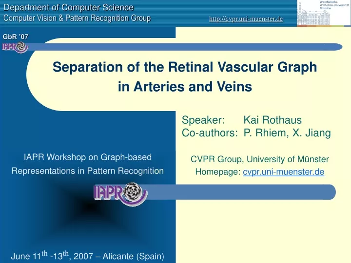
Separation of the Retinal Vascular Graphin Arteries and Veins Speaker: Kai RothausCo-authors: P. Rhiem, X. Jiang CVPR Group, University of Münster Homepage: cvpr.uni-muenster.de
Outline • Introduction • Medical purpose • Image-processing • Method • SAT-problem specification (vessel labelling) • Operations for graph manipulation (edge labelling) • Solving Conflicts • Results • Conclusions and further work Rothaus, Rhiem and Jiang: Separation of the Vascular Graph in Arteries and Veins
Medical Purpose • Why retinal vessel are of interest? • Vessels of retina and brain are conjuct • Only on retina vessels are visible directly • Conclusions on diseases are possible • Anatomy of the eye • Vessels enter the eyeball at the optic disc • Vessels only branch (no reconnection) • Capillars are invisible • Differences of two vessel types on retina: Rothaus, Rhiem and Jiang: Separation of the Vascular Graph in Arteries and Veins
Vessel segmentation • Input: Retinal Image • Output: Binary vessel image • Many segmentation algorithms, based on • Matched-filter • Tracking • Intensity riges or (1st moment deviations) • Curvature (2nd moment deviations) • Special difficulties • Handling of bifurcations and crossings • Central-light reflex • Different vessel width • Wide intensity spectrum • Pathological objects nearby • Mainly, we use hand-segmented images Rothaus, Rhiem and Jiang: Separation of the Vascular Graph in Arteries and Veins
binary vessel image skeleton image vasculature graph Graph-based representation of the vasculature • Input: Binary vessel image • Output: Vasculature graph • Compute the skeleton of the vasculature • Classify skeleton pixel in • End pixel (form vertices of degree 1) • Connection pixel (form edges) • Branching pixel (form vertices of degree 3) • Crossing pixel (form vertices of degree 4) • Construct graph-based representation • Arising Problems: • Segmentation errors could lead to small cycles • Discontinuous segmentation leads to an over-fragmented graph representation • Skeleton of a crossing could lead to two branches Rothaus, Rhiem and Jiang: Separation of the Vascular Graph in Arteries and Veins
a v v a a v a v a v a v v a SAT-Problem Specification (vessel labelling) • Problem: Classify each vessel as artery (a) or vein (v) • Mainly recent approaches are based on local features • Colour, cross-profile, thickness, etc. • Work only good for thick vessels nearby the optic disc • We propose a structure-based approach (on vasculature graph) • Label each vessel segment vi as artery (Li=a) or vein (Li=v) • Formalise anatomical properties of the vasculature: • At branches only edges of the same labelling are involved • At crossings an artery crossing a vein • Construct logical clauses that describe the properties • Cumulate above rules for all vertices and formulate the SAT-problem • Solve this as a CSP (Constraint Search Problem) with AC-3 Rothaus, Rhiem and Jiang: Separation of the Vascular Graph in Arteries and Veins
conflict conflict manuallabel manuallabel The labelling process (AC-3*) • Add the incident vertices of few manually labelled vessel segments in the process queue Q • While Q is not empty • Take the first vertex and corresponding logical rule • Reduce set of labels of the incident vessels consistent to the rule • If there is a conflict try to solve it (details later) • Otherwise add the new vertices to Q • Order of processing the vertices (rules) is important Rothaus, Rhiem and Jiang: Separation of the Vascular Graph in Arteries and Veins
conflict Q={ v6 } Q={ v3,v8 } Q={ v4,v8 } Q={ v8,v7 } Improvement: Introduce an intelligent initial edge labelling to detect split crossings Q={ v7 } Rothaus, Rhiem and Jiang: Separation of the Vascular Graph in Arteries and Veins
vasculature graph edge labelling vessel labelling Operations for graph manipulation (edge labelling) • Segmentation or skeleton errors lead to unsolvable SAT-problem • Graph structure has to be manipulated slightly • Allowed operations should handle: • Split crossings(instead of 1 deg. 4 vertex 2 adjacent deg. 3 vertices) • Missing segments(crossing degenerated to vertex of degree 3) • Falsely detected branches • Falsely detected segments • Instead of manipulating the graph directly we introduce a second order labelling (edge labelling): Rothaus, Rhiem and Jiang: Separation of the Vascular Graph in Arteries and Veins
Steering the labelling process (Belief propagation) • Plausibility weights [0,1] for each vertex • Assign crossing vertex the plausibility 1 - P1(d) • Assign branch vertex the plausibility (with β =max αi) P1(d)+P2(β) - P1(d)P2(β) • Plausibility weights [0,1] for each a/v-labelled vessel • Assign hand-labelled vessels plausibility 1 • During AC-3* algorithm use a multiplicative propagation scheme (with weights of corresponding vertex and edge) • Use weights as heuristic to order Q as priority-queue • Use the average vessel weights to rate labelling results P1(d) P2(β) Rothaus, Rhiem and Jiang: Separation of the Vascular Graph in Arteries and Veins
Initial edge labelling • Decide on plausibility measures P1(d) and P2(β) if a connection edge between to branches is probably a crossing • No false c-label should be introduced • Label edge with c-label iff [ d<3 ]or[ P1(d)<0.75 andP2(β)<P2(30°) ] Confusion matrix on 10 training images Accuracy of >96 % Rothaus, Rhiem and Jiang: Separation of the Vascular Graph in Arteries and Veins
Solving Conflicts • Conflicts cannot been avoided (even not with initial labelling) • Conflicts are basically introduced by cycles in the vascular graph • Topology is responsible for conflicts • Solving-strategy: • Search cycle (vertex set V’), where all vessel labels are defined • Establish edge candidate set E’={ e | e incident to a v in V’ } • Choose a “suitable” n-labelled edge of E’, with minimum weight and change edge label to c (crossing) • Otherwise label the conflict edge with e (end-segment) • Restart the AC-3* algorithm Rothaus, Rhiem and Jiang: Separation of the Vascular Graph in Arteries and Veins
artery (auto.) vene (auto.) artery (man.) vene (man.) Original image Binary image Interactive labelling tool • Requirement: binary vessel image • Physician mark single vessel segments as arteries an veins • Propagation of the manual labelling as far as possible • Solve logical conflicts automatically • If the result is not good enough for the observer, more vessel label could be manually added • Presenting results in two different ways: Rothaus, Rhiem and Jiang: Separation of the Vascular Graph in Arteries and Veins
Results on manually segmented images • STARE data set of A. Hoover et al. image im0082 Rothaus, Rhiem and Jiang: Separation of the Vascular Graph in Arteries and Veins
Discussion results on manual segmentations • Most conflicts could be solved by introducing c-label • Only few conflicts could not been solved • Problematic regions are even hard to been labelled by experts • Normally few hand-labels are necessary Rothaus, Rhiem and Jiang: Separation of the Vascular Graph in Arteries and Veins
Results on automatic segmentations • Method of Soares et al. and test database DRIVE of Staal • High demands on segmentation algorithm:Different vessel width, no gaps in segmentation, low false positive rate, etc. • Some segmentations leads to poorly connected graphs (less rules) Rothaus, Rhiem and Jiang: Separation of the Vascular Graph in Arteries and Veins
Summary and Conclusions • We have developed a method for propagating vessel classification • Requirement is a binary vessel image • Problem is formulated as Constraint Search Problem • Arising conflicts are solved by manipulating graph structure • Interactive environment is developed for physicians • Methods works good for tested image databases • Quality depends strongly on segmentation result • Further works • Statistical foundation of plausibility function • Realise initial labelling with Bayesian classifier • Justify method by comparison with ground-truth data • Enhance conflict solver • Classify strong vessel automatically as artery or vein • Integrate method in a framework for vascular structure analysis Rothaus, Rhiem and Jiang: Separation of the Vascular Graph in Arteries and Veins
Final slide Thank you for your attention! Are there any questions? Rothaus, Rhiem and Jiang: Separation of the Vascular Graph in Arteries and Veins