Download
1 / 97
1.01k likes | 1.26k Views
Microbiology. Prokaryote Architecture. Simple in shape, but genetically and biochemically advanced. General Prokaryote Shapes. Coccus – round or spherical Bacillus – rod shaped Spirila / Vibrio – spiral or twisted, corkscrew, halfmoon. Diplo – groups of 2 Strepto - chains

E N D
Prokaryote Architecture • Simple in shape, but genetically and biochemically advanced
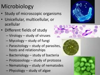
General Prokaryote Shapes • Coccus – round or spherical • Bacillus – rod shaped • Spirila / Vibrio– spiral or twisted, corkscrew, halfmoon
Diplo – groups of 2 • Strepto - chains • Staphlo – grape like clusters
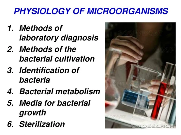
Microscopy • Compound Light Microscope: • Helps to determine cell size and shape • Some internal structures may be seen • Usually need special dye
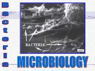
Microscopy • Electron Microscope (to 150,000X): • Transmission (TEM) – helps to see internal cellular features (DNA, cell wall/membrane, ribosomes, etc.) • Scanning (SEM) – helps to see external features (cell surface, envelope, flagella, etc.) TEM – HIV on lymph SEM – RBC’s in clot SEM – E.coli on sm intestines SEM – intestinal tape worm TEM – flu virus
Typical Prokaryotic Components • Cell membrane – selectively permeable barrier separating inside of the cell from its environment • Cell wall – rigid structure surrounding cell membrane • Gives structural support • Protection from lysing (breaking) • Made of peptidoglycan (sugar protein polymer) • Sensitive to PCN
… 3. Ribosomes – combination of RNA & protein • Site of protein synthesis • Sequence of RNA nucleotide bases is used to identify species 4. Chromosome – DNA of cell • Almost always only 1 per cell • May be 2-4 copies in an actively growing cell 5. Inclusions – single structure of molecules of C, P, S, N • Stockpiles of necessary nutrients for future use in metabolism
Flagella - structure that allows cell to be mobile in an aqueous habitat • Classified based on how many flagella
2 Major Groups of Bacteria Gram Positive Bacteria Gram Negative Bacteria About 90% of all bacteria Thin cell wall Small amounts of peptidoglycan Pink • About 10% of all bacteria • Thick cell wall • Contains lots of peptidoglycan • Purple
Prokaryotic Cell Walls Diagrams of the cell wall structure of Gram-negative (left) and Gram-positive bacteria. Key: peptidoglycan layer (yellow); protein (purple); teichoic acid (green); phospholipid ( brown); lipopolysaccharide (orange).
Gram Staining • Simple staining technique used to differentiate the 2 groups of bacteria • Uses the differences in the cell walls of different bacteria • Specific Steps to process: • Heat fix slide • Crystal violet (1 min) • Iodine (1 min) • Alcohol (5-10 secs) • Safranin (1 min)
***Bacteria vary from minutes to years in reproduction time!*** • Microbial growth – increase in the number of cells in a population (group of individuals of the same species) • right conditions must exist. • DNA replication, transcription, and translation have to occur • Proteins, lipids, & polysaccharide synthesis all occur simultaneously
Exponential Growth – population increases in number of cells in a fixed time period (1 → 2→ 4→ 8→ 16 → 32…) • Ideal growing conditions must be present • Steady nutrient supply & space • Unchecked / unlimited growth
Growth Curve of Bacteria **Assumes abundant space, food, and no competition!
Lag (acclimation) Phase – culture is transferred to fresh media • Requires time to adjust for growth to begin (synthesize DNA, enzymes) • Log (exponential) Phase – time of rapid growth • Exponential increase • Unlimited resources, ideal growing conditions
Stationary Phase – log ends as nutrients & space are used up and waste products build up • Balance b/w reproduction and death • Cells began to encounter environmental stress • Lack of water, nutrients, & space • Build up of waste • Changes in oxygen and pH • Death Phase – cells cease metabolism; they become inactive or die due to limiting factors in the environment • Some ‘dead’ cells are alive but enter into “suspended animation” or form spores • Both can grow again
Have the growth curve because we can measure the number of total cells in broth culture (blood, tissue, water) • Microscopy • Spread Plating • Turbidity
Microscopy • Direct cell count by counting the cells within a grid (field) then extrapolating to total volume
Spread Plating • Plate counts after a serial dilution, then count colonies, and extrapolate total volume
A plate count may be done on plates prepared by either the pour plate method or the spread plate method.
Turbidity • Use a spectrophotometer • Use a broth culture in a tube and insert in machine • Amount of light blocked by cells is proportional to the number of cells.
Which method is better? • Best approach is to use turbidity after a plate count • Use 2 methods @ 1st, then turbidity
Nutrition & Metabolism • Catabolism – breakdown of chemicals to release energy • Anabolism – “biosynthesis” – building of larger molecules • Requirements for Growth • Physical • Temperature ( -15ْ C to 125ْC) • pH (-0.05 to 13) • Salt (0% to 30%) • Osmotic pressure • Chemical • micro & macro elements • Oxygen (0-21%)
Temperature • Cold • psychrophiles (cold loving) – organisms that grows best -15ْC up to 15ْC. Don’t grow above 25ْC • Psychrotolerant (cold tolerant) - organisms that grows best >20ْC, but can grow at lower temps • Middle • Mesophile organism that grows best b/w 20ْC • Hot • Thermophiles (hot loving) – organisms that grow best above 45ْC but below 80ْC • Hyperthermophiles (extreme thermophiles) grow best above 80ْC
pH • Acidophile – organisms that grow best below pH of 6 • many foods, such as sauerkraut, pickles, and cheeses are preserved from spoilage by acids produced during fermentation • Neutrophile (neutral pH) – anything b/w 6 & 8 • Where most bacteria grow best • Alkalinophile (basic) – grows best pH above 8
Salt • Halotolerant – tolerant to salt, don’t require salt, but can grow in presence of salt • Halophiles – require some salt for growth (up to 10%) • Extreme halophiles- require at least 10% salt for growth
Osmotic Pressure • Microbes obtain almost all their nutrients in solution from surrounding water • Tonicity • isotonic • hypertonic • hypotonic
Major Elements & Uses in Cells MACRONUTRIENTS • H (8%), O (20%)– H20 • Carbon (C) 50%– major constituent of all macromolecules; uptaken by cells as organic carbon or as CO2 • Nitrogen (N) 14%– major element in proteins and nucleic acids; uptaken as NH3, NO3-, N2, & organic molecules • Phosphorus (P) 3%– major element in ATP, phospholipids, & nucleic acids; uptaken as PO43- • Sulfur (S) 1%– used in amino acids (cysteine, methionine) & vitamins; uptaken as SO42- & HS
MICRONUTRIENTS • Potassium (K+) 1%– transport of small molecules across the cell membrane, helps in enzyme function, & involved in protein synthesis • Sodium (Na+) 1%– can be used by enzymes & in membrane transport, but it is not required by all species • Magnesium (Mg2+) 0.5%– stabilizes DNA and helps in enzyme function, such as DNA polymerase and in ATP productions • Metals (Fe2+, Fe3+, Cu, Zn) – used in electron transport, used by proteins involved in electron transport processes (metabolism)
Can remember these by: CHOPKNS CaFe all except Mg & Na
Oxygen • There are 4-5 different oxygen requirements for bacteria • Obligate aerobe • Facultative aerobe • Microaerophile • Aerotolerant anaerobe • Obligate anaerobe
Obligate aerobe • Require full level of O2 (20-21%) to grow
Facultative aerobes • Grows best in O2, but can grow without O2
Microaerophiles • Grow at O2 levels <20%, but require less
Aerotolerant anaerobes • Don’t require O2, but can grow in presence of O2
Obligate anaerobes • No O2 required, O2 is toxic
Obligate aerobic • Obligate anaerobic • Facultative aerobe • Microaerophile • Aerotolerant
What does all this mean? • You need to know what the organism you are culturing requires for growth so you can study and treat them!!
Methods to Control Growth • Heat Sterilization • Radiation • Filtration • Antimicrobial Agents
Heat Sterilization Sterilization – destruction of all viable life • Incineration (dry heat) – glassware, metal objects • 160ْ – 550ْC • Denatures proteins and makes organic molecules unstable • Takes seconds to hours • Pasteurization (low heat over time) – milk, fluids • 63ْ – 72ْC • Kills up to 99% organisms in milk • 15 seconds – 30 minutes • Autoclave (moist heat) – glassware, metal objects, liquids (sm. Vol.), plastics • 121ْ & 15 psi; pressure is used to increase temp. • Minutes to hours
Radiation • Ionizing radiation (gamma rays) – breaks DNA; disrupts important genes= death • Used for plastics, antibiotics, food • Ultraviolet radiation (ultraviolet rays) – DNA & RNA absorbed and forms strong bonds between thymine; prevents DNA replication • Sterilizes water, air, and surfaces
Filtration • Size of pores or matrix of fibers capture cells while air or fluid passes through