Download
1 / 210
2.18k likes | 3.07k Views
ST220 Concorde Career College. Microbiology. Microbiology. Objectives Define the term microbiology. Provide a timeline of events leading up to the current understanding of microbiology. List and identify the parts of a microscope and understand its use in a laboratory setting. Microbiology.

E N D
ST220 Concorde Career College Microbiology
Microbiology Objectives • Define the term microbiology. • Provide a timeline of events leading up to the current understanding of microbiology. • List and identify the parts of a microscope and understand its use in a laboratory setting.
Microbiology Microbiology – Study of microorganisms Microorganisms – Microscopic organism (plant or animal) Microscopic – Minute; visible only with the aid of a microscope
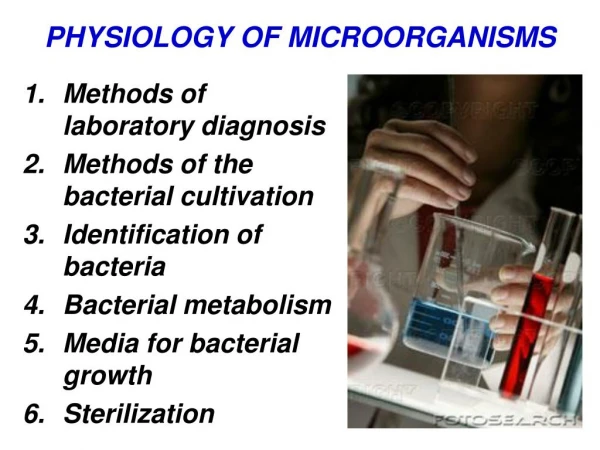
Microbiology Microscope • An optical instrument for making an enlarged image of an object too small to be seen by the naked eye • Consists of a lens, or a combination of lenses • Often provides light enhancement • May be monocular or binocular
Microbiology Microscope Original van Leeuwenhoek microscope
Microbiology Microscope Operating microscope used for neurosurgical procedures
Microbiology Microbiology Timeline • Robert Hooke • 1635-1703 • Cell theory – Proposed the idea that all living things are composed of cells after viewing slices of cork through a microscope in the year 1665. Note: Hooke coined the term cell which is from the Latin word cella meaning "storeroom" or "small container"
Microbiology Microbiology Timeline • Anton van Leeuwenhoek • 1632-1723 • First to observe bacteria with a microscope • Made the first accurate drawings of bacteria and protozoa
Microbiology Microbiology Timeline • Francesco Redi • 1626-1697 • Disproved the theory of abiogenesis or spontaneous generation (it was thought that new living cells could spontaneously arise from nonliving material)
Microbiology Microbiology Timeline • Louis Pasteur • 1822-1895 • Proved the theory of biogenesis (new living cells can only arise from preexisting living cells) • Developed the “germ theory of disease” • Promoted hand washing and a clean environment to reduce the spread of disease in hospitals • Developed several vaccines (anthrax, cholera, rabies) • Developed "pasteurization," a method of destroying harmful microbes in perishable food products using heat, without destroying the food
Microbiology Microbiology Timeline • Edward Jenner • 1749-1823 • Developed smallpox vaccination • Father of Immunology
Microbiology Microbiology Timeline • Ignaz Semmelweis • 1818-1865 • Correlated frequent handwashing in the obstetrics ward with lower rates of infection (puerperal fever) Note: the term puerpera refers to a woman who has just given birth
Microbiology Microbiology Timeline • Joseph Lister • 1827-1912 • Recognized the significance of Pasteur’s findings and established the first principles of asepsis and the practice of sterile technique • Used carbolic acid (phenol) to treat surgical wounds and dressings • Wore clean gown and gloves for each surgical procedure • Boiled instruments between surgical procedures
Microbiology Microbiology Timeline • Robert Koch • 1843-1910 • Proved Pasteur’s germ theory • Developed pure culture techniques still used today
Microbiology Microbiology Timeline • Hans Gram • 1853-1938 • Developed staining methods (Gram’s stain) utilized to identify various species of bacteria
Microbiology Organism Identification • Staining – Involves application of a colored dye (called a stain) to an organism to enhance visualization and allow identification of a specific organism. • Gram Stain • Acid-fast Stain
Microbiology Organism Identification • Gram Stain • Organism is stained with blue/purple dye • Weak iodine solution is added to promote colorfastness • Slide is washed with alcohol • If blue/purple dye remains the organism is Gram positive • If blue/purple dye is removed the organism is Gram negative and is then stained with pink/red dye to enhance visibility
Positive or Negative? Anthrax rods in CSF Pseudomonas aeruginosa
Microbiology Organism Identification • Acid-fast Stain • Organism is stained with red dye • Smear is treated with acid • Most organisms quickly loose the red color when treated with acid • Those that remain colored (e.g., Mycobacterium tuberculosis) are called acid-fast
Acid-fast Stain Mycobacteria paratuberculosis in bovine ileum
Microbiology Organism Identification • Culturing – Growing cells in enriched media • Fermentation – Action of organism on sugars • Observation – Reaction to test chemicals • Inoculation – Injection and observation of test animals • Immunology – Antigen-antibody reaction
Microbiology Microscope Types • Compound Light Microscope • Dark Field Microscope • Phase Contrast Microscope • Fluorescence Microscope • Electron Microscope
Microbiology Compound Light Microscope • Also called “bright field” • Two lens system • First lens located in the objective near the specimen • Second lens in the eyepiece • Light source
Microbiology Dark Field Microscope • Similar to bright field microscope – contains a light condenser with an opaque disc that blocks direct light; allowing peripheral light to enter • Utilized when microbes cannot be stained • Effective for viewing motility when microbes are suspended in liquid (wet mount)
Microbiology Phase Contrast Microscope • Light condenser contains ring shaped diaphragm that allows light of various brightness (phases) to pass through – highlighting the internal structures of the cell
Microbiology Fluorescence Microscope • Utilized ultraviolet light to visualize fluorescent microbes or those stained with fluorochromatic dye • Especially useful in identification of rabies and syphilis
Microbiology Electron Microscope • Utilizes a beam of electrons in place of light which improves resolution • Utilizes an electromagnetic lens in place of a glass lens which focuses the electron beam onto the specimen • Scanning electron microscope is pictured
Electron Microscope Pollen
Microbiology Microscope Components • Eyepiece • Body tube • Arm • Revolving nosepiece • Objectives • Stage with clips • Focus adjustment knobs • Diaphragm • Light source • Base
Microbiology Microscope – Care and Handling • When carrying the microscope hold the arm with one hand and support the base with the other • Never touch the lenses • Use only lens paper to clean the lenses • When finished with the microscope • Rotate the nosepiece to the low power objective • Lower the nosepiece down to the stage • Secure the cord • Replace the dust cover
Microbiology Objectives • List and identify the components of a cell and describe the function of each. • List and define fluid movement concepts. • List the major classifications of organisms.
Microbiology • Cell Theory • Anton von Leeuwenhoek • Observed pond water under microscope • Noted organisms that he called “animalcules” • Principles of modern cell theory • Cells are the smallest complete living things (basic unit of organization of all organisms) • All organisms are composed of one or more cells in which all life processes occur • Cells arise only from preexisting cells through the process of cell division. • All existing cells are descendants of the first cells formed in history.
Microbiology • Cell Structure • Cell membrane • Cytoplasm • Nucleus • Nuclear membrane • Nucleoplasm • Chromatin • Nucleolus • Mitochondria • Lysosomes • ER – endoplasmic reticulum (rough, smooth) • Golgi apparatus • Ribosomes