Download
1 / 23
240 likes | 473 Views
Protein Structure Database for Structural Genomics Group. M.S. Thesis Defense. Jessica Lau December 13, 2004. Bioinformatics is Analysis of biological data: gene expression, DNA sequence, protein sequence. Data mining and management of biological information through database systems.

E N D
Protein Structure Database for Structural Genomics Group M.S. Thesis Defense Jessica Lau December 13, 2004
Bioinformatics is • Analysis of biological data: gene expression, DNA sequence, protein sequence. • Data mining and management of biological information through database systems. • At the Northeast Structural Genomics Consortium, database management systems play a large role in its daily operation • Data collection and mining of experimental results • Track target progress – status milestones • Exchange information with rest of the world • My thesis presents work in database management systems at the NESG. • Part 1: ZebaView • Part 2: Worm Structure Gallery • Part 3: Prototype of NESG Structure Gallery
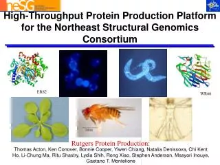
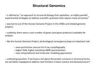
Zebaview is the official target list of the Northeast Structural Genomics Consortium • Display summary table of NESG targets. • Status milestones • Protein properties: DNA and protein sequences, molecular weight, isoelectric point • New targets are curated and then uploaded to SPiNE. • 11,284 targets from 88 organisms.
Family View NESG Families • Unfolded • Membrane • Core 50 • Nf-kB
Selected Cloned Expressed Soluble Purified X-ray or NMR data collection In PDB • 4,418 targets cloned • 141 structures • 3.4% successful targets Target Summary Statistics
GO, Cellular Localization, and SignalP • Search for targets that have • any of the three GO ontologies defined • no GO ontologies defined at all 116 NESG structures do not have Molecular Function defined
LOCTarget • Secretory proteins require formation of disulfide bonds • Oxidative Folding needed for proper native folding • 2,132 “Extracellular” NESG targets Bovine ribonuclease A has four disulfide bonds to stabalize its 3-D structure. Mahesh Narayan, et al. (2000) Acc. Chem. Res., 33 (11), 805 -812.
SignalP • mRNA are translated with signal peptide for cellular localization • Peptide is cleaved upon destination • SignalP predicts cleavage of signal peptide • Removal of signal peptide gives proper native fold Lodish et al. Molecular Cell Biology 4th edition, Figure 7.1 (2000)
Caenorhabditis elegans • Widely studied model organism • 2-3 weeks life span, small size (1.5-mm-long), ease of laboratory cultivation, transparent body • Small genome, yet has complex organ systems similar to higher organisms: digestive, excretory, neuromuscular, reproductive systems Donald Riddle et al, C. elegans II (1997) Altun Z F and Hall DH. , Atlas of C. elegans Anatomy, Wormatlas (2002-2004)
System Components • 22,653 C. elegans proteins • 42 experimentally determined • 4 are from NESG • 24 homology models • 14 are from NESG • 960 C. elegans proteins potentially modeled • Uniprot: Pfam domain, Gene name, ORF name • PDB Coordinates • Structure Validation Report • Sequence similarities to proteins in PDB
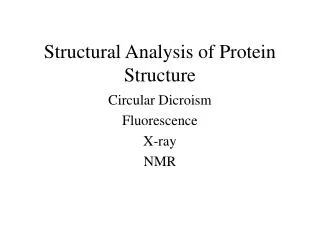
Protein Structure Validation Software • Suite of quality validation software • PROCHECK • Quality of experimental data • Distribution of φ, ψ angles in Ramachandran plot • MolProbity Clashscore • Number of H atom clashes per 1,000 atoms • With respect to a set of scores from 129 high resolution X-ray crystal structures • < 500 residues, of resolution <= 1.80 Å, R-factor <= 0.25 and R-free <= 0.28; Bahattacharya, A et al. to be published
Homology Modeling Automatically (HOMA) • Algorithm based on alignment between query and template sequences. • Regions of conserved residues forms a set of constraints for modeling • Sequence identity of 40% or more • Good quality template
Search • Search for C. elegans proteins in local database. • Keyword: “Ubiquitin” in any field Results: 72 C. elegans proteins 2 Experimentally determined structures 1 Homology model 11 Potential models Results: 152 C. elegans proteins 2 Experimentally determined structures 1 Homology model 19 Potential models
System Architecture • Java, Tomcat, MySQL, Perl. • Three-tier architecture • Client: Web browser • Application: JSP, Logic components, Data access components • Data: MySQL
Structure files submitted by automated pipeline • ADIT integrated with SPiNE for uniform format • PSVS and images automatically generated • Structure information from PSVS directly into SPiNE • Archives structure files. • Structure files submitted by individual groups • Structure information is entered into SPiNE manually • Manually run PSVS and MolScript
Downloads • Structure Validation Report • Structure related files • Atomic coordinates • NMR constraints • NMR peak lists • Chemical shifts • Structure factor • Annotation • Functional annotation provided by other NESG members • Uniprot • PDB coordinates file • Reusing Java components from Worm Structure Gallery
Future Direction • Enhance ZebaView performance to handle increased load and functionalities • Integrate annotation from other protein and structure databases. • Make modules available for other java-based applications within structural genomics. • Develop a gallery for other organisms: yeast, fruit fly, human • Continue specifications for the new NESG Structure Gallery
Thank You Advisor: Dr. Gaetano Montelione Thanks to everyone at the Protein NMR lab and NESG! Aneerban Bhattacharya John Everett All the scientists who solved the structures!