Download
1 / 46
460 likes | 468 Views
Explore the detailed anatomy and functions of the male and female reproductive systems, including the structures, processes, and hormones involved in producing and nurturing sex cells. Learn how these systems play vital roles in human reproduction.

E N D
Reproductive System • Male and female system are a connected series of organs and glands • Produce and nurture sex cells • Transport the sex cells to fertilization site • Secrete hormones vital for development and maintenance of secondary sex characteristics • Regulate reproductive physiology
Male Reproductive Organs • Produce and maintain male sex cells, sperm cells • Transport these cells and supporting fluid to outside • Secrete male sex hormones
Male Reproductive Organs • Primary sex organ: 2 testes • Make sperm cells and male sex hormones • Accessory sex organs: internal and external reproductive organs
Testes • Ovoid structures about 5cm in length and 3 cm in diameter • Both testes are within the saclike cavity of the scrotum
Testes Structure • Composed of lobules separated by connective tissue and filled with seminiferous tubules • Epithelium lining the seminiferous tubules produces sperm cells • Interstitial cells produce male sex hormones
Sperm Formation • Epithelium lining the seminiferous tubules include: • Supporting cells: support and nourish spermatogenic cells • Spermatogenic cells: give rise to sperm cells • Sperm cells consists of a head, midpiece, and tail
Spermatogenesis • Give rise to sperm • Meiosis reduces the number of chromosomes in sperm cells by ½ • 46 23 chromosomes • Produces 4 sperm cells for each primary spermatocyte
Male Internal Accessory Organs • Epididymis: a tightly coiled tube that leads into the vas deferens • Stores and nourishes immature sperm cells and promotes their maturation
Male Internal Accessory Organs • Vas deferens: muscular tube • Passes through the inguinal canal and enters the abdominal cavity • Courses medially into the pelvic cavity and end behind the urinary bladder • Fuses with the duct from the seminal vesicle to form the ejaculatory duct
Male Internal Accessory Organs • Seminal Vesicle: a saclike structure attached to the vas deferens • Secretes an alkaline fluid that contain nutrients, such as fructose, prostaglandins
Male Internal Accessory Organs • Prostate Gland: surrounds the urethra just inferior to the urinary bladder • Secretes a thin, milky fluid that neutralizes the pH of semen and acidic secretions of the vagina
Male Internal Accessory Organs • Bulbourethral glands: two small structures inferior to the prostate gland • Secrete a fluid that lubricates the penis in preparation for sexual intercourse • Semen: consists of sperm cells and secretions of the seminal vesicles, prostate gland, and bulbourethral glands • This fluid is slightly alkaline and contains nutrients and prostaglandins
Male Internal Accessory Organs • Sperm Cell: located in semen • Swim and have the ability to fertilize egg cells once in female reproductive tract
Male External Reproductive Organs • Scrotum: a pouch of skin and subcutaneous tissue • Encloses the testes • Penis: specialized to become erect for insertion into the vagina during sexual intercourse • During erection, the vascular spaces within the tissue become engorged with blood • Semen moves along the reproductive tract as smooth muscle in the walls of the tubular structures contracts by reflex
Male Hormones • Hypothalamic and Pituitary Hormones • Male body remains reproductively immature until the hypothalamus releases gonadotropin-releasing hormone (GnRH) • Stimulates the anterior pituitary gland to release gonadotropin • Follicle-stimulating hormone (FSH) stimulates spermatogenesis • Luteinizing hormone (LH) stimulates interstitial cells to produce male sex hormones
Male Sex Hormones • Androgens • Production increases rapidly at puberty • Testosterone: most important • Stimulates development of male reproductive organs • Male sexual characteristics
Regulation of Male Sex Hormones • A negative feedback mechanism regulates testosterone concentration • Rising testosterone concentration inhibits the hypothalamus and reduces the anterior pituitary’s secretion of gonadotropins • As testosterone concentrations falls, the hypothalamus signals the anterior pituitary to secrete gonadotropins • Testosterone concentrations remain stable from day to day
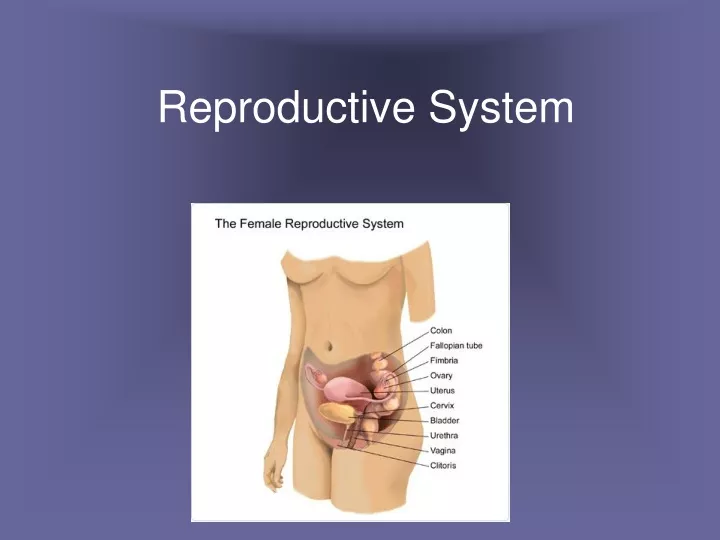
Female Reproductive System • Produce and maintain the female sex cells • Egg cells or ova • Transport these cells to the site of fertilization • Provide a favorable environment for a developing offspring • Move offspring to the outside • Produce female sex hormones • Primary Female Sex Organs: two ovaries
Ovaries • Two ovaries: solid, ovoid structures • 3.5 cm long, 2 cm wide, and 1 cm thick • Lie in the shallow depressions in the lateral wall of the pelvic cavity
Ovary Structure • Subdivided into a medulla and a cortex • Medulla: connective tissue, blood vessels, lymphatic vessels, and nerves • Cortex: ovarian follicles and is covered by cuboidal epithelium
Preimordial Follicles • During prenatal development, groups of cells in the ovarian cortex form millions of preimordial follicles • Each contains a primary oocyte and a layer of follicular cells • Primary oocyte begins meiosis • Process stalls until puberty • The number of oocytes steadily declines throughout a female’s life
Oogenesis • At puberty, some oocytes are stimulated to continue meiosis • Primary oocyte secondary oocyte • Chromosome number is cut in half • 46 23 • Fertilization of a secondary oocyte produces a zygote
Follicle Maturation • At puberty, initiated by FSH • Generally, one follicle matures at a time • Maturation: • oocyte enlarges • Follicular cells multiply • Fluid filled cavity forms
Ovulation • The release of an oocyte from an ovary • A rupturing follicle releases a oocyte • After ovulation, the oocyte is drawn into the opening of the uterine tube
Internal Accessory Organs • Uterine tubes • the end of each tube expands • Margins bears irregular extensions • Ciliated cells that line the tube and peristaltic contractions in the wall of the tube help transport the egg cell down the uterine tube
Internal Accessory Organs • Uterus: receives the embryo &sustains it • Wall includes the endometrium, myometrium, and perimetrium • Vagina: receives the erect penis, coveys uterine secretions to the outside, and provides an open channel for the fetus during birth
External Accessory Organs • Labia Majora: rounded folds of adipose tissue and skin • Upper ends form a rounded elevation over the symphysis pubis • Labia minora: are flatted, longitudinal folds between the libia majora • Well supplied with blood vessels
External Accessory Organs • Clitoris: a small projection at the anterior end of the vulva • Corresponds to the penis • Composed of two columns of erectile tissue • Vestibule: space between the labia minora • Vestibule glands secrete mucus
Female Hormones • Sex cell maturation • Development & maintained of female secondary sexual characteristics • Glands • Hypothalamus • Anterior pituitary gland • Ovaries
Female Hormones • Most important: estrogen & progesterone • Estrogen: develops and maintains most female secondary sex characteristics • Progesterone: changes in the uterus
Female Reproductive Cycle • FSH initiates a menstrual cycle by stimulating follicle maturation • Maturing follicular cells secrete estrogen • Maintains secondary sexual traits • Thickening of the uterine lining • Anterior Pituitary: secretes large amount of LH • Triggers ovulation
Female Reproductive Cycle • Following ovulation, follicular cells give rise to the corpus letuem • Secretes progesterone • Causes the uterine lining to become more vascular and glandular • If an oocyte is not fertilized, the corpus luteum begins to degenerate • As estrogen and progesterone concentrations decrease, the uterine lining disintegrates • Menstrual flow
Female Reproductive Cycle • During the cycle, estrogen and progesterone inhibit the release of LH and FSH • As estrogen and progesterone decline, anterior pituitary secretes FSH and LH again • Stimulate a new menstrual cycle
Menopause • Termination of menstrual cycle due to the aging of the ovaries • Reduced estrogen concentrations and lack of progesterone may cause regressive changes in female secondary sex characteristics
Mammary Glands • Located in the subcutaneous tissue of the anterior thorax • Composed of lobes that contain ducts and glands • Dense connective and adipose tissue separates the lobes
Mammary Glands • Ovarian hormones stimulates female breast development • Alveolar glands and ducts enlarge • Fat deposits around and within the breasts