Download
1 / 49
1.17k likes | 4.05k Views
Opportunistic Mycoses & Miscellaneous Mycoses. III MBBS Dr Ekta Chourasia Department of Microbiology. Opportunistic Systemic Mycoses. Produced by relatively non-pathogenic or contaminant fungi in a host whose immunological defense mechanisms are weakened by

E N D
Opportunistic Mycoses & Miscellaneous Mycoses III MBBS Dr Ekta Chourasia Department of Microbiology
Opportunistic Systemic Mycoses • Produced by relatively non-pathogenic or contaminant fungi in a host whose immunological defense mechanisms are weakened by Endogenous causes - cancer, leukemia Exogenous causes - immuno suppressive therapy, AIDS. • Important cause of morbidity & mortality in hospitalized patients. Dr Ekta, Microbiology
Opportunistic Mycoses • Includes - Candidiasis - Cryptococcosis - Aspergillosis - Zygomycosis / Mucormycosis - Pneumocystosis - Penicilliosis - Miscellaneous opp. mycoses. Dr Ekta, Microbiology
CRYPTOCOCCOSIS • Fungal disease caused by an encapsulated yeast Cryptococcus neoformans, pathogenic to man & animals. • 2nd most common fungal infection after candidiasis in HIV - infected individuals. • Isolated from pigeon nests, droppings, old buildings & nitrogenous soil - Creatine favour the growth. Dr Ekta, Microbiology
Virulence factors • Capsule – Inhibits phagocytosis. • Melanin production by the enzyme phenol oxidase. L- DOPA Melanin Antioxidant - protects the organism from intracellular killing by phagocytes Dr Ekta, Microbiology
Pathogenesis • Infection occurs by inhalation, but sometimesthrough skin or mucosa. • Weakening of immune system leads to reactivation & dissemination to CNS and other sites. • Visceral forms simulate tuberculosis & cancer clinically. • Cutaneous form varies form small ulcers to granulomas. Dr Ekta, Microbiology
Clinical Features • Seen in HIV +ve patients when CD4+ count falls below 200 cells / mm3 • Extra pulmonary cryptococcosis is one of the AIDS – defining disease. • Clinical types : • Pulmonary • Extrapulmonary – CNS, viscera, bones & skin Cryptococcal meningitis is the most serious type of infection, resembles TB and is often seen in AIDS patients. Dr Ekta, Microbiology
Laboratory Diagnosis • Specimen – Serum, CSF, body fluids Direct examination • Wet mount: India ink - budding yeast cells 5-20, with a distinct halo • Gram’s stain - Gram +ve budding yeast cells Dr Ekta, Microbiology
Fungal Culture & Serology • SDA without actidione - highly mucoid, cream to buff colored. • Birdseed (Niger seed) agar – selective media - Brown colored colonies due to melanin production. • Crypto LA test – Ag detection in Serum, CSF & Urine - Titer > 18 is significant. Dr Ekta, Microbiology
Treatment and Prophylaxis • Antifungals - AMB: Drug of choice - Flucytosine • Vaccine - conjugate vaccine developed (capsule component linked to TT) Dr Ekta, Microbiology
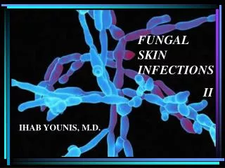
ASPERGILLOSIS • Systemic infection in immunocompromised as well as immunocompetent individuals • Infection occurs by - Inhalation of conidia - Direct entry through wounds or during surgery. • Imp. species – A. niger, A. fumigatus, A. flavus Dr Ekta, Microbiology
Clinical types • Pulmonary Aspergillosis 3 categories depending upon whether the host is atopic or immunocompromised. • Allergic aspergillosis 1. Aspergillus asthma - Atopic individuals - Inhalation of spores Dr Ekta, Microbiology
2. Allergic bronchopulmonary aspergillosis(ABPA) - Repeated & heavy exposure to spores - Breathlessness, fever & malaise 3. Obstructive aspergillosis - Plugs of entangled mycelia & mucus block segments of lung tissue & even entire lobe. - Productive cough: contains aspergillus hyphae. Dr Ekta, Microbiology
B. Aspergilloma • Non invasive • Colonization in a pre- existing cavity (tubercular) • Compact mass of fungal mycelia surrounded by dense fibrous walls : FUNGUS BALL • Usually solitary 8-10 cm C. Invasive Aspergillosis • Important cause of morbidity & mortality • May disseminate to kidneys & brain. Dr Ekta, Microbiology
Extrapulmonary Aspergillosis– CNS, Paranasal sinus, skin, endocardium (in prior cardiac surgery) • Miscellaneous forms - Eye (oculomycosis) - Ear (otomycosis) - Nails (onychomycosis) Dr Ekta, Microbiology
Laboratory Diagnosis • Common lab contaminant – hence repeated isolation from specimen is mandatory. • Specimen – sputum, BAL, tracheobronchial biopsy. Direct Examination • Wet mount – 10% KOH, CFW - hyaline, septate hypha, 3-6 with acute angled (45º) branching. Dr Ekta, Microbiology
Fungal Culture • Grow easily & quickly on routine media. • SDA without actidione. • Identification of species by induction of sporulation on following media : - Czapek Dox agar - 2% malt extract agar • Colony characteristics & the morphology help in identifying the species. Dr Ekta, Microbiology
Aspergillus – Cultural characteristics A.niger A.fumigatus A.flavus
MUCORMYCOSIS • Invasive disease caused by lower fungi – Zygomycetes • Found in food items, soil & air. • Common lab contaminants • Reproduction • Asexual – Sporangiospores within sporangium. • Sexual – single, dark thick walled spores called Zygospores. Dr Ekta, Microbiology
Introduction • Includes following genera - Mucor - Rhizopus distinguished on the - Absidia basis of morphology. - Rhizomucor • Usually occurs in diabetic patients with ketoacidosis. * high glucose & acidotic condition favours their growth. Dr Ekta, Microbiology
Clinical types 1° infection occurs in URT or nose by inhalation • Rhinocerebral– commonest, fulminant type Nasal mucosa – Turbinate bones - Paranasal sinus - Orbit, Palate & Brain • Pulmonary • Cutaneous • Disseminated – lungs, kidney, brain, heart & GIT. Dr Ekta, Microbiology
Laboratory Diagnosis Specimen – nasal discharge, sputum, biopsy Direct Examination • KOH – irregular broad, aseptate ribbon like hyphae with wide angle (90º)branching at irregular intervals. • Special stains – must - HE, GMS Dr Ekta, Microbiology
Fungal Culture • Cottony, dense & floccose colony. • LPCB mount – for microscopic details & species identification. Dr Ekta, Microbiology
MUCOR ABSIDIA RHIZOPUS Dr Ekta, Microbiology
Treatment & Prophylaxis • A life threatening condition • 4 concomitant approaches : • Rapid correction of underlying predisposing conditions like diabetic ketoacidosis. • Surgical debridement of necrotizing tissue. • Antifungals (not azoles) • Adjunctive therapy with hyperbaric oxygen. Ideal treatment = surgical debridement + I.V. AMB Dr Ekta, Microbiology
PENICILLOSIS • Caused by Penicillium marneffei, a dimorphic facultative intracellular fungi. • Only species of genus Penicillium which causes infection. • 3rd most common AIDS - related opportunistic infection after TB & Cryptococcosis. Dr Ekta, Microbiology
Epidemiology • Route of transmission - Inhalation of conidia - Ingestion (eating rats, China) - Direct inoculation of skin. • Bamboo rat harbors P. marneffei in their internal organs. • Isolated from - feces - soil samples from their burrows. Dr Ekta, Microbiology
Pathogenesis & Pathology • RES is the 10 site of infection. • 3 histologic patterns of disease: • Granulomatous – granulomas • Suppurative – multiple abscesses - lungs, skin & subcutaneous tissue of immunocompetent individuals. • Necrotizing - immunocompromised pt. Dr Ekta, Microbiology
Laboratory diagnosis • Clinical diagnosis difficult as symptoms are very similar to other fungal pathogens like H.capsulatum • Presumptive diagnosis - yeast forms with cross- walls in biopsy. - CD4+count < 50 cells /mm3 • Definitive diagnosis : direct demonstration & isolation of organisms in culture Dr Ekta, Microbiology
Direct Examination • Wrights’s, Giemsa stain of skin, biopsies – septate yeast like cells. • Tissues – PAS, GMS • Immunohistochemical assay • Peripheral blood smears – AIDS patient. Dr Ekta, Microbiology
Fungal Culture • Isolated from blood, skin, BM, sputum, LNs, pleural fluid, urine & BAL • SDA & BA at 250C : woolly pigmented colonies, reverse is bright rose color. • Microscopy:- Hyaline, short, septate hyphae with branching. - Brush like conidiophores bearing conidia. Dr Ekta, Microbiology
Culture • At 370 C - SDA, BHIA, 5% sheep BA & Pine’s medium Colony – white chalk like LPCB – pleomorphic yeast cells with transverse septa. • M to Y conversion differentiates from other species of Penicillium. • Treatment - Amphotericin B & Itraconozole Dr Ekta, Microbiology
PNEUMOCYSTOSIS • Infection by inhalation of droplet nuclei – leads to interstitial cell pneumonia, primarily involves alveoli. • Caused by Pneumocystis carinii, a unicellular eukaryote. • Initially it was considered as a protozoa but now considered as a fungi - shares biological features of both groups fungi & protozoa. Dr Ekta, Microbiology
Features of Pneumocystis carinii • Stained with the fungal stains like GMS. • Does not grow on fungal culture media but requires tissue culture or cell lines. • Susceptible to anti-protozoan agents like Pentamidine & TMP-SMZ. • Insensitive to antifungals because of lack of ergosterols. • Produces chitin. • Cyst wall ultrastructure similar to Ascomycetes. • Hence classified as “Atypical” fungus. Dr Ekta, Microbiology
Life Cycle • Divided into 3 stages : - Trophozoite (asexual): fill the alveoli - Cyst (sexual) - Sporozoite (Intracystic body) • The transition phase between trophic & cystic stage is called Precyst(SPOROCYST) Dr Ekta, Microbiology
Clinical features • Incubation period : 4 to 8 weeks. • Pulmonary manifestations – non productive cough, dyspnoea, fever, 20 infections, cyanosis (late sign) • Extrapulmonary manifestations – in advance HIV infection - LNs, BM, spleen, liver, stomach, small intestine, pancreas, thyroid & eyes. - CNS (late complication of AIDS) Dr Ekta, Microbiology
Radiodiagnosis • ‘Ground – glass’ OR Honey comb like appearance – classical finding in PCP. Laboratory diagnosis • CD4+ count < 200 cells / mm3 • Clinical specimen – sputum, BAL or lung biopsy. Dr Ekta, Microbiology
Direct Examination • Selective stains – GMS, Toluidine blue. • Lung biopsy - frothy edema fluid in alveoli. • Immunofluorescence Dr Ekta, Microbiology
Culture • Cannot be cultured on fungal media. • Requires tissue culture : • A- 549, a continuous cell line derived from human lung adenocarcinoma cells. • Human embryonic lung fibroblasts (WI- 38) Dr Ekta, Microbiology
Treatment & Prophylaxis • Combination of TMP & SMZ I.V. – drug of choice • Pentamidine Isothionate I.V. – who cannot tolerate TMP- SMZ. • Dapsone Dr Ekta, Microbiology
Miscellaneous Opportunistic Fungi • Recently established fungal agents. • Includes – Trichosporon - Geotrichum - Rhodotorula - Sacchromyces (Baker’s yeast) Dr Ekta, Microbiology
Miscellaneous Mycoses Oculomycoses • Fungal disease of eye. Can be : • Mycotic keratitis – follows corneal trauma, overuse of corticosteroids. • Endophthalmitis • Infections of ocular adnexa • Causative agents – Aspergillus, Fusarium, C. albicans • Treatment : Local – AMB, Nystatin Dr Ekta, Microbiology
A picture of a Gram stain of scrapings from a corneal ulcer. This was from a farmer who had a piece of vegetable matter embedded in his cornea. The culture grew a pure culture of Aspergillus fumigatus. Dr Ekta, Microbiology
Otomycoses • External ear. • Usually caused by Aspergillus species. • Secondary to bacterial infection, injury or excessive accumulation of cerumen (wax). Dr Ekta, Microbiology
Mycotic Poisoning • 2 types : • Mycetism – fungus which is eaten, itself causes toxic effects. - GI disease, dermatitis or death. e.g. Psilocybe species • Mycotoxicosis – food contaminated by fungal toxins. - “Aflatoxin” produced by A.flavus. Dr Ekta, Microbiology
ANTIFUNGAL AGENTS • Ergosterol – present in the cytoplasmic membrane of only fungi. - most important site of action of most antifungals. • Classification - Antifungal antibiotics - Synthetic antifungals - Miscellaneous antifungals Dr Ekta, Microbiology
A. Antifungal antibiotics • Polyene Antibiotics – • Interferes with sterol synthesis leading to disruption of cell. • Produced by Streptomyces e.g. Amphotericin B, Nystatin, Hamycin • Other antibiotics – Griseofulvin • Produced by Penicillium & Actinomadura • Superficial fungal infections. Dr Ekta, Microbiology
B. Synthetic Antifungals • Thiocarbamates – Tolnaftate • Allylamines & Benzylamines – Terbinafine, Naftifine • Azoles - Inhibits cytochrome P 450 dependent C14 demethylation in the synthesis of ergosterol • Imidazoles – Ketoconazole, Clotrimazole, Econazole, Miconazole • Triazoles – Fluconazole, Itraconazole Dr Ekta, Microbiology
C. Miscellaneous Antifungal Agents • Flucytosine • Whitfield’s ointment • Selenium sulfide • Potassium iodide • Gentian violet paint Dr Ekta, Microbiology