Download
1 / 41
430 likes | 676 Views
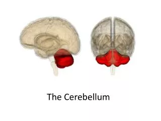
The Cerebellum. DR. S. H. KHAN. Position. Lies behind the fourth ventricle, medulla and pons Occupies posterior cranial fossa Largest part of Hind brain Coordinates voluntary movements of the body. Cerebellum. External features.

E N D
The Cerebellum DR. S. H. KHAN
Position • Lies behind the fourth ventricle, medulla and pons • Occupies posterior cranial fossa • Largest part of Hind brain • Coordinates voluntary movements of the body Cerebellum
External features Consists of twocerebellar hemispheres united in the midline by the vermis Vermis Hemisphere Superior view Inferior view
External features Tonsil of cerebellum two elevated masses on inferior surface of hemispheral portion just nearby foramen magnum Tonsil
Fissures & Lobes • Two deep fissures • Primary fissure • Posterolateral fissure • Three lobes • Flocculonodular lobe flocculus and nodule • Anterior lobe • Middle lobe (Posterior lobe) Corpus of cerebellar
Fissures & Lobes Anterior lobe corpus of cerebellar Primary fissure Middle lobe Flocculonodular lobe Posterolateral fissure
Internal structures Gray matter • Cerebellar cortex • Intracerebellar nuclei (embedded in white matter) • Dentate nucleus • Fastigial nucleus • Interposed nucleus • Emboliform nucleus • Globose nucleus White matter • medullary center
Internal structures Fastigial nucleus Cerebellar cortex Globose nucleus Dentate nucleus Emboliform nucleus medullary center
Three functional divisions • Archicerebellum • Flocculonodular lobe • Lingula of vermis • Paleocerebellum • Vermis and • intermediate zone • Neocerebellum • Lateral zone Intermediate zone Vermis Lateral zone Flocculonodular lobe
Functions of Cerebellum • Controls the same side of the body • Regulates and coordinates voluntary movements of the body • Keep the movements smooth, balanced and accurate • Planning of sequential movement • Controls muscle tone, posture and equilibrium
Injury : Cerebellar syndrome • Hypotonia • Postural changes & alteration of Gait • Ataxia(disturbance of voluntary movements) Intention tremor • Dysdiadochokinesia ( inability to perform alternating movements regularly & rapidly) • Nystagmus (abnormal movements of eyeballs) • Dysarthria (disorder of speech)
Connections of cerebellum • Cerebellum is connected to the brainstem by 3 paired bundles of nerve fibers ( Peduncle) Three peduncles • Inferior cerebellar peduncle -connect with medulla and spinal cord, -contain both afferent and efferent fibers • Middle cerebellar peduncle -connect with pons, -contain afferent fibers • Superior cerebellar peduncle -connect with midbrain, -contain mostly efferent fibers
Position of Diencephalon • Position: Lies between midbrian and cerebrum, almost entirely surrounded by cerebral hemisphere
Subdivisions of Diencephalon • Thalamus (Dorsal thalamus) • Metathalamus • Epithalamus • Subthalamus (Ventral thalamus) • Hypothalamus
Dorsal thalamus interthalamic adhesion External features • A large egg-shaped nucleus (mass of gray matter) • Situated in the lateral wall of 3rd ventricle and in the floor of the lateral ventricle • Anterior end called anterior thalamic tubercle, • Posterior end called pulvinar • The right & left thalami are joined by interthalamic adhesion
Structure of Thalamus • White matter -Stratum zonale- thin layer covers the superior surface -External medullary lamina- covers the lateral surface -Internal medullary lamina- Y shaped vertical sheet, divides the gray matter into 3 parts • Gray matter -Anterior thalamic nuclei - Medial thalamic nuclei - Lateral thalamic nuclei - Intralaminar nuclei
Subdivision of dorsal thalamus Three main parts -divided by internal medullary lamina (Y) • Anterior part (Anterior nuclear group) • Medial part (Medial nuclear group) • Lateral part (Lateral nuclear group) Int. medullary lamina Ant part Med. part Lat. part
internal medullary lamina Med. nuclear group Lat. posterior Ant. nuclear group Lat. dorsal Pulvinar Ventral anterior Medial geniculate body (MGN) Ventral intermediate Ventral posterior nucleus (VP) Lateralgeniculate body (LGN) Ventral posterolateral (VPL) Ventral posteromedial (VPM )
Function of Thalamus • Great integrating center for sensory impulses • Receives sensory information of all types, except smell • Receives profuse connections from all parts of cerebral cortex, cerebellum and corpus striatum. • Receives exteroceptive (pain, temp. touch), proprioceptive (muscles, joints) and visceral informations Injury • Sensory loss • Thalamic syndrome
Metathalamus • Lies posteroinferior to thalamus • Consists of medial geniculate body lateral geniculate body
Metathalamus Lateralgeniculate body (LGN) Medial geniculate body (MGN) Metathalamus
Metathalamus • Medial geniculate body (MGN) • Relay station of audition • Receive fibers from inferior colliculus • Projects to auditory area via acoustic radiation • Lateralgeniculate body(LGN) • Relay station of vision • Receive fibers from optic tract • Projects to visual area via optic radiation
Epithalamus Includes • Thalamic medullary stria • Habenular nucleus • Habenular trigone • Habenular commissure • Pineal body • posterior commissure Pineal body
Subthalamus • Situated between thalamus and midbrain • Contain subthalamic nucleus, parts of red nucleus and substantia nigra
Position-lies below the thalamus Forms the floor & inf. Part of lat. Wall of third ventricle Boundaries Superiorly: hypothalamic sulcus Inferiorly: optic chiasma tuber cinereum Infundibulum mamillary body Anterior: lamina terminalis Posterior: continues with midbrain tegmentum Lateral : internal capsule Hypothalamus
hypothalamic sulcus optic chiasma lamina terminalis mamillary body Infundibulum
Subdivisions • Preoptic region • Optic region • Tuberal region • Mamillary region
Important nuclei • Optic region • Supraoptic nucleus -produce antidiuretic hormone (ADH/ vasopressin ) • Paraventricular nucleus- produce oxytocin • Tuberal region • Infundibular nucleus • Ventromedial nucleus • Dorsomedial nucleus • Mamillary region • Mamillary nucleus • Posterior hypothalamic nucleus
Paraventricular nucleus Paraventriculohypophyeal tract Supraoptic nucleus Mamillary nucleus Supraopticohypophyseal tract arcuate nucleus tuberoinfundibular tract Infundibulum of pituitary anterior lobe of hypophsis posterior lobe of hypophysis
Hypothalamus --connection • Connects with limbic system • Connects with brainstem and spinal cord • Connects with dorsal thalamus • Connects with hypophysis
Hypothalamus --connection • Supraoptic nucleus supraoptic nucleus (ADH) →supraopticohypophyseal tract →posterior lobe of hypophysis • Paraventricular nucleus paraventicular nucleus (oxytocin) →paraventriculohypophyseal tract→posterior lobe of hypophysis
Paraventricular nucleus Paraventriculohypophyseal tract Supraoptic nucleus Supraopticohypophyseal tract Inferior hypophyseal a. posterior lobe of hypophysis Hypophyseal v.
Hypothalamus Function • Regulates functions of neuro-endocrine system -Secrets ADH & Oxytocine, which then transported to posterior pituitary gland by nervous stimulation -By forming Releasing/ Inhibitory hormones, controls Ant. Pituitary gland to secret hormones ( through minute vessels of Hypothalamic-Hypophyseal system) • Autonomic nervous system -Controls CVS, Respiratory, Alimentary functions