Download
1 / 75
810 likes | 921 Views
Integumentary System. Skin. Functions. Protects underlying tissues from dehydration Regulate body temperature Manufacture vitamin D Site for nerve endings Temporary storage of fat, glucose, water and salts Protects body from foreign invaders (1 st line of defense). Functions continued.

E N D
Integumentary System Skin
Functions • Protects underlying tissues from dehydration • Regulate body temperature • Manufacture vitamin D • Site for nerve endings • Temporary storage of fat, glucose, water and salts • Protects body from foreign invaders (1st line of defense)
Functions continued • Screen out harmful Ultraviolet radiation • Absorb certain drugs and chemical substances • Two or more kinds of tissues grouped together and perform specialized functions constitute an organ. • Largest organ (150 lb = 20 sq. ft.) • Used for sensation
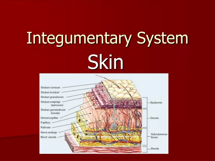
Basic layers • Epidermis • Outermost covering • Made of stratified squamous epithelium • Dermis • Inner layer • Thicker than the epidermis • Includes connective tissue, epithelial tissue, smooth muscle, nervous tissue, and blood • Subcutaneous • Beneath the dermis • Loose connective tissue, predominantly adipose • NOT a true layer of skin
Epidermis • Four distinct cell types and five layers • Thickness varies • Thinnest is the eyelids • Thickest is on the palms of the hands and soles of the feet • Surface layer (stratum corneum) consists of dead cells rich in keratin
Keratin is a protein that renders the skin dry and provides a waterproof covering • Keratin also serves as a barrier against ultraviolet light, bacteria, abrasions, and some chemicals.
Epidermal Cells • Keratinocytes • Comprise most of the epidermis and produce the protein keratin • Merkel cells • Sensory receptors • Melanocytes • Make melanin, protects the skin against UV, skin color is due largely to melanin. • Langerhans cell • Macrophages which defend the skin
The Largest Organ • The skin of a 150-lb person would cover 20 square feet
Epidermal layers • Stratum corneum (top layer) • Dead, flat, keratinized cells; slightly acidic • Stratum lucidum • Found only on the palms of the hands and soles of the feet • Layer appears clear • Stratum granulosum • Keratinization process begins and cells begin to die
Epidermal layers continued • Stratum spinosum • 8 to 10 cell layers thick • Melanocytes • Keratinocytes • Langerhans cells
Epidermal layers • Stratum germinativum/basale • Undergoes cell division • Deepest epidermal layer • Mostly keratinocytes • Grow upward and become part of the superficial layer • Melanocytes and merkel cells are found here
Cells of the _______________ are non-living cells and are rubbed away by friction. • Stratum corneum • Stratum Lucidum • Stratum granulosum • Stratum spinosum
Which layer of epidermis can only be found in the thickened skin of the palms and soles? • stratum granulosum • stratum lucidum • stratum basale • stratum corneum
The cells of the epidermis that reproduce are in the • keratin. • stratum corneum. • stratum basale. • stratum granulosum.
Dermis • Thicker • Inner layer • Matted masses of connective tissue, collagen tissue bands, elastic fibers, nerve endings, muscles, hair follicles, oil, sweat glands, and fat cells • Sensory nerves end in nerve receptors • Pressure receptors are deeper in the dermis
Dermis Layers • Papillary layer - The most superior layer of the dermis. Characterized by papillae, fingerlike projections that intermix with the epidermis. Consists primarily of loose connective tissue.
Dermis Layers Reticular layer - The reticular layer of the dermis (RD) consists of dense irregular connective tissue. Gives the skin its overall strength and elasticity, as well as housing other important epithelial derived structures such as glands and hair follicles.
Subcutaneous or Hypodermal layer • Under the dermis • It is not a true part of the integumentary system • Loose connective tissue/adipose • Contains one-half of the body’s stored fat • Hypodermis layer attaches the integumentary system to the surface muscles underneath • Site of injections • Innermost layer
Accessory Structures of the skin • Hair • Nails • Sudoriferous (sweat) glands • Sebaceous (oil) glands • Ducts
Hair Follicle • Follicle extends from the surface into the dermis and contains the hair root. • Hair shaft extends from the skin. • Hair is composed of dead epidermal cells. • Pigment is determined by epidermal melanocytes. Dark hair has more of the brownish-black eumelanin, white blonde hair and red hair have more pheomelanin.
Sudoriferous/Sweat Glands • Sudoriferous - any of the glands in the skin that secrete perspiration • Eccrine glands – most abundant, common on the forehead, neck, and back where they produce sweat. • Apocrine Sweat Gland - Sweat glands that release their secretions into hair follicles. Produce a highly individual sexual scent
Sebaceous Glands • Sebaceous gland - Glands in the skin of mammals that secretes an oily substance called sebum. • In humans, sebaceous glands are primarily found in association with hair follicles but also occur in hairless areas of the skin, except for the palms of the hand and soles of the feet. • Helps to keep the hair and skin soft, pliable, and waterproof
Glands • Exocrine Glands – secrete products into ducts. (example: Sweat, Salivary) • Endocrine Glands – do not secrete products into a duct. (example: Pituitary Gland, Pancreas) • Duct: a circumscribed channel leading from an exocrine gland or organ.
Rally Robin • Lefty – Tell your partner about 1 accessory structure in the skin • Righty – Tell your partner about 1 other accessory structure in the skin • 30 seconds – repeat until time runs out
Skin Color • All people have same # of melanocytes • Differences result from the amount of melanin produced by melanocytes • The amount is determined by our genes • Sunlight, UV light, & X rays stimulate production • Well-oxygenated blood = bright red, so skin looks pinkish • Poorly oxygenated blood = dark red, skin looks bluish (cyanosis)
Relationship to microorganisms • Handwashing • Minimum time is 10 to 30 seconds • Sing “Row, row, row your boat” x 2 • Contact with blood or infectious materials • First wash hands, apply gloves before exposure, remove gloves, wash hands again.
Burns • 1st degree – burn injuring only the epidermis. Produce redness, pain, and minor swelling • 2nd degree – destroys the epidermis as well as some dermis. Produce blisters, severe pain, and redness. • 3rd degree – destroys the epidermis, the dermis, and the accessory structures. The surface appears dry and can look waxy white, leathery, brown, or charred
The type of burn that destroys the epidermis and dermis layer is • 2nd degree • 3rd degree • Yo Momma jokes • 1st degree
Disorders • Acne – combo of dead skin cells, sebum, and bacteria plugging up hair follicle • Athlete’s foot – fungus • Dermatitis – inflammation of the skin, rash • Eczema – noncontagious inflammatory skin disease, skin becomes dry, red, itchy, and scaly • Impetigo – contagious skin disease, in babies and young children. Ruptured vesicles, caused by staph/strep organism
Acne • Def - occurs when your hair follicles become plugged with oil and dead skin cells. • Causes – overproduction of sebum (oil) • Irreg. shedding of dead skin cells • Bacteria • Worsened by hormones, meds, diet • S&S – whiteheads, blackheads, pimples, papules, nodules, & cysts on the face, neck, back, chest, & shoulders
Acne • Treatment – • Topical treatment/lotion (OTC/Rx) • Antibiotics • Oral contraceptives • Light/laser therapy • Chemical peels/dermabrasion
Acne • Prevention • Wash 2x daily • Acne cream • Avoid heavy makeup • Remove makeup before bed • Loose clothing • Shower after exercising
World’s Largest Pimple • http://www.youtube.com/watch?v=7OjDqGC3YBw
Athlete’s Foot • Def – (tineapedis) fungal infection usually between the toes • S&S – • scaly red rash • Itching, stinging, burning • blisters or ulcers • Cause • Damp & warm socks & shooes • humid conditions favor the organisms' growth • Contagious • Contact w/ infected person or surfaces (towels, floors, shoes)
Athlete’s Foot • Treatment – • Prevention -
Psoriasis – inflammatory skin disease, dry reddish patches covered with silvery-white scales. Usually found on the elbows, knees, skins, scalp, and lower back. • Ringworm – highly contagious fungal infection. • Urticaria or hives – itching welts, response to allergen