Download
1 / 104
1.55k likes | 3.35k Views
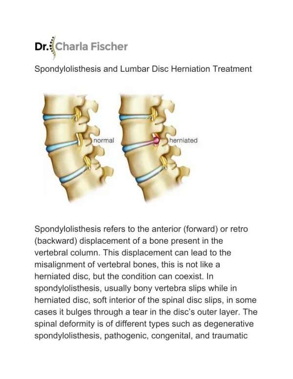
Spondylolisthesis. Jwalant S. Mehta MS( Orth ), D ( Orth ), MCh ( Orth ), FRCS ( Tr & Orth ) Consultant Spine Surgeon, ABMU Health Board. Outline of the talk. Classification Natural history Patho -physiology Treatment rationale Cases. Spondyl Olisthesis.

E N D
Spondylolisthesis Jwalant S. Mehta MS(Orth), D (Orth), MCh (Orth), FRCS (Tr & Orth) Consultant Spine Surgeon, ABMU Health Board
Outline of the talk Classification Natural history Patho-physiology Treatment rationale Cases
SpondylOlisthesis 1741 Nicholas Andry: hollow back 1782Herbiniaux Belgian obstetrician 1854Kilian slow displacement ‘Spondylolisthesis’ 1855 Roberts: No slip if arch intact
Newman & Stone JBJS Br 1963; 45: 39 - 59
Meyerdings grades Low Grade High Grade I II III IV V
Important in grades III – V Slip angle
Pelvic incidence Pelvic tilt Sacral slope PI = PT + SS
Low PT High SS High PT Low SS
Relevance of pelvic measures PI quantifies the pelvic shape Pelvic morphology and spino-pelvic balance are abnormal in spondylolisthesis
Hook and catch Hook: Pedicle Pars inter-articularis Inferior process of the cephalad level Catch: Superior process of the caudal level
Pathophysiology Dysplastic pathway Traumatic pathway
Dysplastic pathway Traumatic pathway Repetitive cyclic loads (sports) Weakness in the hook & catch mechanism Body weight transmitted through weak zone Stress fracture of a Normal pars Hard cortical pars pre-disposes to fatigue fracture and non-union Soft tissue restraints: plastic deformation Growth plate overloaded Predisposes to a vertical subluxation
Dysplastic changes Contributes to the mechanics of progression, but not causation Proximal sacral rounding Trapezoidal L5 Vertical sacrum Junctional kyphosis Compensatory hyper-lordosis
proximal sacral rounding Yue Spine 2005
Discal over-loading Alternative loading pathway Haher Spine 1994 Both the pathways lead to ↑ shear loads, axial loads remaining constant Premature disc degeneration
The pain generators: Back pain Leg pain is the most common symptom Moller Spine 2000 • Chronic muscle spasm (protective): • ‘painful’ pars • Annular tears • Root compression / traction
The pain generators: Leg pain L5 compression / traction Abnormal motion Facet joint arthrosis Pars scar The disc above far-lateral
Clinical evaluation: history • Symptoms: • Back pain • Leg pain • Neurology • Severity • Activities of daily living
Clinical evaluation: examination Range and rhythm of trunk motion Neurology Sagittal alignment & gait
Sagittal alignment Stance Gait Head over pelvis Hips and knees
Imaging • Erect radiographs: • AP • Lateral (to include the hips) • MRI; CT • Occasionally: SPECT; Dynamic radiographs; Discography
Purpose of imaging Disc degeneration (MRI / CT) Facet joint orientation, tropism, degeneration (MRI / CT) Pelvic and spinal measures (Erect xrays)
Disc degeneration: MRI Grade II Grade III Grade IV Grade V Grade I Pfirrmann et al Spine 2001
Facet joints: orientation & tropism Don JSDT 2008 Wang Spine 2009 Boden JBJS Am 1996 Vanharanta Spine 1993 • Mean facet joint angle: Sagittal: anterior forces • Tropism R –L: asymmetric loads • Mild < 5° • Moderate 7° – 15° • Severe > 15°
Facet degeneration: cartilage Grogan et al AJNR 1997 Uniformly thick layer Focal erosions Areas of deficiency with exposed bone Cartilage absent except traces
Facet degeneration: sub-chondral sclerosis Grogan et al AJNR 1997 Thin layer of cortical bone Focal thickening Thick < ½ of the surface Dense cortical bone > ½ of the surface
Facet degeneration: osteophytes Grogan et al AJNR 1997 No osteophyte Small Moderate Large
Centre for Spinal Studies and Surgery Nottingham Severe Spinal Stenosis
Wiltse classification:III. Degenerative • Instability phase: Kirkaldy Willis • Posterior elements are intact • L45; F >M • Disc: • degeneration, • ↓ height • Facets: • Tropism • Abnormal sagittal orientation • Facetal arthritis; subluxation
Natural history: genetics Albanese JPO 1982 Wynne-Davies JBJS Br 1979 Roche JBJS Am 1952 Stewart JBJS Am 1953 15 – 70% 1st degree relatives Lysis commoner in boys Slips commoner in girls Eskimos 25% (arch defects)
Natural history: ‘the slip’ Bentley Spine 2003 15% of persons with a pars lesion During the growth spurt Minimal change after 16 y No pain during progression
Extent of the problem Seitsalo JBJS Br 1990 Danielson Spine 1991 Frennerd JPO 1991 Seitsalo Spine 1991 Most are asymptomatic 90% slips at initial presentation do not progress
Progression risk Ohmori JBJS Br 1995 Muschik JPO 1996 > 20 y: more stable, less symptomatic, less likely to progress High level of athletic activity, no effect on progression Association with back pain ‘weak’
The risk of progression in the young adult: disc degeneration
Clinical Growth yrs (9 – 15) Girls > Boys Back pain Postural or gait abn Radiographic Type 1 (dysplastic) Vertical sacrum >50 % slip Increasing slip angle Instability on flex/ext views Risk factors for slip progression in spondyolisthesis(Hensinger 1989)
Adolescents III+: likely to progress I, II after mid-adolescence: unlikely to progress Natural history of progression
Always consider first……………….everytime! Improvement likely if back > leg pain Isthmic / degnerative with leg pain: improvement less likely Investigate / treat osteopaenia Non-operative treatment
Stop aggravating activities Gradual mobilisation Trunk strengthening Period of bracing Non-operative treatment: Paediatric