Download
1 / 26
330 likes | 1.04k Views
Trauma– Blunt Abdominal Trauma. Douglas M. Maurer, DO, MPH. Learning Objectives. Recognize and respond appropriately to a patient with hemorrhagic shock Assess via bedside methods the source of hemorrhage

E N D
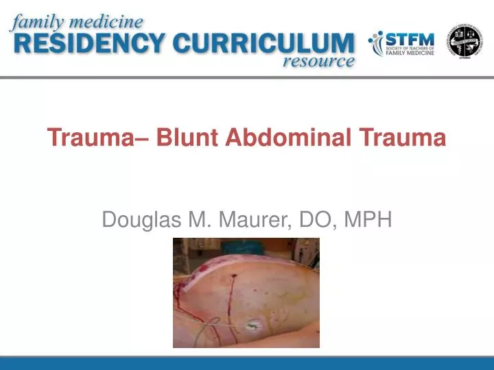
Trauma– Blunt Abdominal Trauma Douglas M. Maurer, DO, MPH
Learning Objectives Recognize and respond appropriately to a patient with hemorrhagic shock Assess via bedside methods the source of hemorrhage Respond appropriately to evidence of intra-abdominal hemorrhage with regards to initial management and disposition
Introduction Blunt abdominal trauma is common. Unknown history, distracting injuries, and altered mental status make these patients difficult to diagnose and manage. Victims frequently have both abdominal and extraabdominalinjuries. Family physicians need to be able to recognize and treat hemorrhagic shock.
Recognition of Hemorrhagic Shock Shock: oxygen delivery < tissue demands Treatment must restore tissue perfusion not just blood pressure Shock does NOT SBP < 90mmHg Recognition includes: mechanism of injury, patient’s appearance, vitals, level of mentation, peripheral perfusion and urine output Clinical parameters should be coupled with objective markers of tissue perfusion--serum lactate, base deficit, etc.
Practical Diagnosis of Shock Perform a targeted physical examination Diagnostic testing should include chest radiography, pelvis radiography, and bedside ultrasound Objective serum makers of tissue perfusion (serum lactate or base deficit) Point of care H/H, send CBC, type/cross DON’T delay resuscitation for lab results
6 Steps to Treat Hemorrhagic Shock Step 1: Effectively manage the airway and optimize oxygenation. Step 2: Identify and control immediate threats to central perfusion. Step 3: Identify and address severe intracranial injuries. Step 4: Identify and control other potentially life-threatening thoracic and abdominal injuries. Step 5: Identify and control potentially limb-threatening injuries. Step 6: Identify and treat noncritical injuries.
Treatment of Hemorrhagic Shock • Obtain immediate type and crossmatch for 6-8 units of blood • Massive transfusion defined as > 10 U of PRBCs in 24 hrs • Consider use of PRBC to platelet to FFP ratio of 1:1:1 • May result in decreased need for blood products • Give calcium to prevent citrate toxicity
Assessing for Sources of Hemorrhage • Chest radiography: • Tension pneumothorax? Massive hemothorax? Aortic injury? • Pelvis radiography: • Pelvic ring disruption? • Focused Assessment with Sonographyfor Trauma (FAST): • Pneumo/hemothorax? Hemopericardium? Hemoperitoneum? • If positive, then emergency laparotomy. • If negative, continue resuscitation, treat other causes.
FAST Facts • Reliably identifies 200-250ml of intraperitonealfluid • Cannot reliably evaluate retroperitoneum/hollow viscous injury • Sensitivity/specificity: 75%/98%, NPV: 94%; 86-97% accurate • Performed using a curvilinear 2.5 or 3.5 MHz probe
FAST Views Cardiac: parasternal or subxiphoid, hepatocardiacinterface, pericardial space. RUQ: hepatorenal interface (Morrison’s Pouch), diaphragm, inferior pole of kidney. LUQ: splenorenal interface, diaphragm, inferior pole of kidney, inferior tip of spleen. Suprapubic: outline of bladder, silhouette of uterus (females).
FAST Algorithm • Unstable patient: + FAST = OR. • Stable pt: + FAST = abdominal CT. • Stable pt, low mechanism of injury: - FAST = observation, serial exams. • CT is the “Gold Standard”.
What About Diagnostic Peritoneal Aspiration (DPA)? • Can be performed if - FAST in blunt abdominal trauma. • If DPA +, then emergency laparotomy. • If DPA -, then seek and treat other sources. • Perform serial abdominal exams. • Perform serial FAST exams. • If patient stabilizes, then CT. • Get surgery involved!
Indications for Emergency Laparotomy Peritonism Free air under the diaphragm Significant gastrointestinal hemorrhage Hypotension with +FAST scan or + DPA Do NOT keep trauma patients if you lack resources to care for them!
Summary • Recognize and treat hemorrhagic shock aggressively with blood products • Assess for hemorrhage with bedside methods: CXR, pelvis, and FAST • Unstable patient: + FAST = OR. • Stable pt: + FAST = abdominal CT. • Stable pt, low mechanism of injury: - FAST = observation, serial exams.
References • Puskarich MA. Initial evaluation and management of blunt abdominal trauma in adults. In: UpToDate, Hockberger RS, Moreira ME (Ed), UpToDate, Waltham, MA, 2012. • Nickson C. “Trauma! Blunt abdominal trauma decision making.” Weblog entry. Life in the Fastlane Blog. http://lifeinthefastlane.com/2012/03/trauma-tribulation-023/ • Eastern Association for the Surgery of Trauma Guidelines Workgroup. Evaluation of blunt abdominal trauma. 2010 Edition. Chicago, IL. http://www.east.org/resources/treatment-guidelines/category/trauma • American College of Surgeons. ATLS Textbook, 9th Edition. 1 September 2012.
Simulation Training Assessment Tool (STAT)– Blunt Abdominal Trauma Douglas M. Maurer, DO, MPH, FAAFP
Simulation Training Assessment Tool (STAT)– Blunt Abdominal Trauma Learning Objectives: 1. Recognize and respond appropriately to a patient with hemorrhagic shock. 2. Assess via bedside methods the source of hemorrhage. 3. Respond appropriately to evidence of intra-abdominal hemorrhage with regards to initial management and disposition. Date: 1 May 2013 Instructor(s): Clark, Maurer, Cuda Learner(s): ME = Meets Expectations; NI = Needs Improvement, M = Milestones (see debriefing sheet)