Download
1 / 35
460 likes | 1.14k Views
Signaling by Serine/ Threonine Kinase Receptors. Major Classes of Protein Ser/ Thr Kinases (not a complete list). 2 nd -Messenger-dependent protein kinases cAMP -dependent protein kinase cGMP -dependent protein kinase Ca 2+ /CaM-dependent protein kinases Protein kinase C

E N D
Major Classes of Protein Ser/ThrKinases(not a complete list) • 2nd-Messenger-dependent protein kinases cAMP-dependent protein kinase cGMP-dependent protein kinase Ca2+/CaM-dependent protein kinases Protein kinase C • 2nd-Messenger-independent protein kinases MAPK kinase cascade and target kinases Raf, MEK kinases, MEKs, SEKs, ERKs, JNKs, SAPKs, RSKs
Major Classes of Protein Ser/ThrKinases (cont’d) • Cyclin dependent kinases (CDKs) and CDK regulating kinases Cdc-2, CAK, CAK kinase • GPCR kinases GRK2 (βARK1), GRK3 (βARK2), GRK5, GRK6 • P21-activated kinases (PAK) • Kinases involved in cytoskeletal organization and development ROCK/Rho kinase • Transmembrane receptor protein ser/thrkinases TGFβ receptor protein kinase • Casein kinases CK1, CK2
Smad 6/7 are inhibitory - they act as decoys and occupy the sites that would normally be utilized by other activating Smads. Other proteins such as noggin and chordin bind BMPs and inhibit their ability to activate receptors Follistatin inhibits signaling via activins
Ser/Thr Protein Kinases 2 Major Types: • 2nd-messenger-dependent protein kinases. • 2nd-messenger-independent protein kinases.
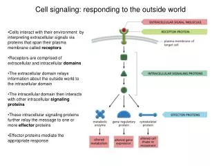
2nd-Messenger-Dependent Protein Kinases All kinases in this category share common design: Several functional domains that can reside on the same pp chain or on separate ones. Each kinase has a catalytic domain (intrinsically active), which is kept inactive by a regulatory domain. Regulatory domain have auto-inhibitory regions and binding sites for 2nd messengers. Interaction with the 2nd messenger dissociates the auto-inhibitory site from the cat domain dis-inhibiting it. Additional regions of the kinases may be responsible for oligomerization or for targeting the kinases to distinct cellular locations.
PKA The function of cAMP Targeting PKA (cyclic-AMP-dependent protein kinase A)
Schematic view of the different domains of 2nd-messenger- dependent protein kinases. The different kinases contain a regulatory domain that encodes specialized functional domains and a conserved cat domain kept inactive by the auto-inhibitory region (black).
PKA • Binding affinities of reg subunits to cAMP: RIIβ < RIIα < RIα < RIβ Necessary for decoding the cAMP signals that differ in intensity and duration: PKAI activated transiently by weak cAMP signals PKAII activated by persistent and strong cAMP levels. RIα may be protective against proteolytic degradation (as in, say, when one of the other subunits are removed (gene targeting)).
Ca2+/CaM kinases(next slide) (A) Domains and holoenzyme structure of CaMKII. (B) Mechanism of autophosphorylation. Thr286. Displacement of the autoinhibitory domain by calmodulin exposes T286, which can then be phosphorylated if its proximate neighbor is active. (C) Trapping of calmodulin to the CaMKII subunits increases the P(autophosphorylation) during successive Ca2+ spikes at high frequency.
Ca2+/CaM kinases Autophosphorylation of Thr286/287 has 2 consequences: • Calmodulin remains bound to the phosphorylated subunit for extended periods of time even at low [Ca2+] (trapped state) because the autophosphorylation greatly decreases the calmodulin dissociation. • Autophosphorylatedα and β subunits are rendered Ca/CaM-independent (autonomous), but still retain substantial kinase activity. Both these consequences (calmodulin trapping and autonomy) allow the phosphorylatedkinase subunits to remain active beyond the limited duration of a Ca2+ spike. Transgenic mice carrying Thr286A1a point mutation in αCaMKII do not exhibit LTP. NMDAR subunits (NR2B) remain active even after dissociation of Ca2+/CaM.
PKC Family consists of at least 10 structurally related phospholipid-dependent protein kinases. Most are expressed in brain. All isozymes grouped into 3 subclasses according to their regulatory properties (conferred by specific domains of the protein): “Conventional”: (cPKCs; α, βI, βII, γ) are regulated by Ca2+, DAG, or phorbol esters and PS. “Novel”: (nPKCs; δ, ε, θ, η) activated by DAG or phorbol esters and PS, but are Ca2+-independent. “Atypical”: (aPKC; μ) unresponsive to DAG, Ca2+, or phorbol esters.
PKC Refer to Schematic view of PKC (earlier slide): Single polypeptide chain = N-term regulatory domain and C-term cat (kinase) domain. Regulatory: auto-inhibitory or pseudo-substrate site and 1-2 membrane targeting motifs (C1 and C2). C1 binds phorbol esters and DAG. C2 binds acidic phospholipids and Ca2+ (in the cPKCs).
PKC Regulation of PKC isozymes: requires removal of the auto-inhibitory pseudo-substrate from the active site (high specific binding of DAG and PS to the C1 and C2 domains) conformational Δ This allows translocation of PKCs from cytoplasm to the membrane enzyme is maximally activated. But, both phosphorylation of PKCs and specific protein-protein interactions are also important for their activation.
PKC One critical protein-protein interaction is with PDK-1: PDK-1 phosphorylates PKC auto-phosphorylation of 2 additional residues. Necessary for the newly synthesized PKC isozymes to achieve catalytically competent conformation. Inactive conformation: binds AKAPs(anchoring kinase associated proteins) and 14-3-3. Active conformation: RACKs(receptors for activated C kinase) Both kinds of binding proteins aid in shuttling PKC to specific compartments.
PKG A dimer of 2 identical subunits. Compared to the other kinases, very limited cellular distribution. The 2nd messenger, cGMP, very limited in its actions.
2nd-Messenger-Independent Protein Kinases MAPK Cascade. See next slide for repertoire of neuronal GEFs that convey on Ras and Rap the extranuclear signals leading to MAPK activation and gene expresion.
Neuronal Substrates of Kinases Neurotransmitter Release E.g., PKA and CaMKIIphosphorylatesynapsin I in the N-terminus. CaMKIIphosphorylatessynapsin I in the C-terminus. ERKsphosphorylatesynapsin I in both N- and C-terminus.
Neuronal Substrates of Kinases Ligand-gated ion channels and K+ channels E.g., CaMKIIphosphorylatesAMPARs (LTP). E.g., PKA phosphorylates GluR1 subunits .incrP(opening) of AMPARs. E.g. PKC phosphorylates GluR2 subunits differentially regulates its interaction with PDZ domain proteins, GRIP1 and PICK1. E.g., PKC, PKA, CaMKII, and ERK all phosphorylate Kv4.2 (VG K+ channel). E.g., PKC and PKA phosphorylate the Kir1 channel.
Neuronal Substrates of KinasesTranscription FactorsMany Different KinasesPhosphorylate CREB CREB – end-point of several signaling pathways
Role of the Kinases in Synaptic Transmission and Cross-Talk Between the Different kinase Pathways
Role of Kinases in Synaptic Plasticity • PKA • CaMKII • PKC • MAPKs
Post-synaptic Signaling by Glutamate Release in an Excitatory Synapse
Serine ThreoninePhosphatases • Critical for signaling and regulating complex neuronal functions (e.g., synaptic plasticity) activated by phosphorylation. • Arise from different genes with no homology. • Impossible to i.d. consensus sequences for their specificity of substrate recognition. • The same phosphatases can dephos substrates that have been phos by different kinases. • A limited number of multifunctional phosphatases (PP1 and PP2B) account for most phosphatase activity.
Serine ThreoninePhosphatases Protein Phosphatase 2B (PP2B, Calcineurin) • Ca/CaM-dependent phosphatase. • Highly enriched in brain. • Heterodimer [A (60 kDal) and B (19 kDal ) subunits]. • A subunit: N-term cat domain and C-term reg domain. • B subunit: CaCaM binding domain. • PP2B activated by low [Ca2+].
Serine ThreoninePhosphatases Protein Phosphatase 2B (PP2B, Calcineurin) • At least 3 PP2B binding proteins have been identified as inhibitors of the enzyme: CAIN, AKAP 79, and FKBP12. • E.g., FKBP12 promotes the assoc of PP2B to ryanodine and IP3 receptors allowing PP2B to regulate [Ca2+]freecyto. • E.g., AKAP 79 binds both PP2B and PKA, bringing them close together for the regulation of receptors and ion channels.
Serine ThreoninePhosphatases Protein Phosphatase 2B (PP2B, Calcineurin) • PP2B expressed highly in hipp: in dendritic spines, but is virtually absent in glia and interneurons. • PP2B is specific for CREB’s ser133. • In mice over-expressing the auto-inhibitory domain of PP2B, LTP, at subsaturatingtetanization, but not saturating conditions, was enhanced • Inhibition of PP2B facilitates LTP formation.
Serine ThreoninePhosphatases Protein Phosphatase 1 (PP1) • Several subunit catalytic isoforms, α, β, γ1, and γ2, interacts with other proteins in subcellular targeting. • E.g., PP1 binds Yotiao, which, in turn, also binds NR1 receptors and the RII reg subunit of PKA so that PP1 and PKA activity are target to the NMDARs. • PP1 activity can be regulated in various ways, including direct inhibition:
Serine ThreoninePhosphatases Protein Phosphatase 1 (PP1) • There are at least 4 known PP1 inhibitory proteins: Inhibitor-1 (I1), inhibitor-2 (I2), dopamine, and cAMP-regulated phosphoprotein (DARP-32), and nuc inhibitor (NIPP1). • PP1 activity is differentially regulated by the phosphorylation of proteins: Phos of I1 and DARP-32 inhibits PP1 activity; Phos of I2 and NIPP1 activate PP1.
Gating Mechanism for PP1 Regulation PKA PP2B ATP ADP ADP ATP I1 (T35) and DARP-32 (T34) cAMP pathway activation would phosphorylate and activate DARP32 Ca2+ pathways activation would dephosphorylate and inactivate DARP-32 Signals