Download
1 / 73
730 likes | 743 Views
The skeletal system, composed of bones, ligaments, tendons, and cartilages, plays crucial roles in support, protection, movement, mineral storage, and blood cell formation. Learn about bone classification, structure, bone cells, and functions of different bone types in this comprehensive guide to the skeletal system.

E N D
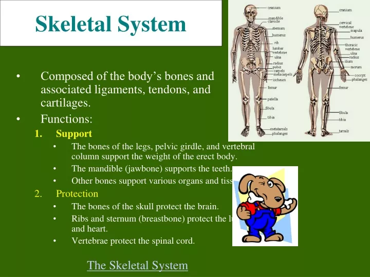
Skeletal System • Composed of the body’s bones and associated ligaments, tendons, and cartilages. • Functions: • Support • The bones of the legs, pelvic girdle, and vertebral column support the weight of the erect body. • The mandible (jawbone) supports the teeth. • Other bones support various organs and tissues. • Protection • The bones of the skull protect the brain. • Ribs and sternum (breastbone) protect the lungs and heart. • Vertebrae protect the spinal cord. The Skeletal System
Skeletal System • Functions: • Movement • Skeletal muscles use the bones as levers to move the body. • Reservoir for minerals and adipose tissue • 99% of the body’s calcium is stored in bone. • 85% of the body’s phosphorous is stored in bone. • Adipose tissue is found in the marrow of certain bones. • Hemopoiesis • A.k.a. blood cell formation. • All blood cells are made in the marrow of certain bones.
Bone Classification • There are 206 named bones in the human body. • Each belongs to one of 2 large groups: • Axial skeleton • Forms long axis of the body. • Includes the bones of the skull, vertebral column, and rib cage. • These bones are involved in protection, support, and carrying other body parts. • Appendicular skeleton • Bones of upper & lower limbs and the girdles (shoulder bones and hip bones) that attach them to the axial skeleton. • Involved in locomotion and manipulation of the environment.
Bone Classification Femur • 4 types of bones: • Long Bones • Much longer than they are wide. • All bones of the limbs except for the patella (kneecap), the bones of the wrist and ankle. • Consists of a shaft plus 2 expanded ends. • Your finger bones are long bones even though they’re very short – how can this be? • Short Bones • Roughly cube shaped. • Bones of the wrist and the ankle. Skeletal System Carpal Bones
Bone Classification • Types of bones: • Flat Bones • Thin, flattened, and usually a bit curved. • Scapulae, sternum, (shoulder blades), ribs and most bones of the skull. • Irregular Bones • Have weird shapes that fit none of the 3 previous classes. • Vertebrae, hip bones, 2 skull bones ( sphenoid and the ethmoid bones). Sternum Sphenoid Bone
Bone Structure • Bones are organs. Thus, they’re composed of multiple tissue types. Bones are composed of: • Bone tissue (a.k.a. osseous tissue). • Fibrous connective tissue. • Cartilage. • Vascular tissue. • Lymphatic tissue. • Adipose tissue. • Nervous tissue. Microscopic Structure of the Bone
All bones consist of a dense, solid outer layer known as compact bone and an inner layer of spongy bone – a honeycomb of flat, needle-like projections called trabeculae. Above: Note the relationship btwn the compact and spongy bone. Below:Close up of spongy bone. Skeletal System
Note the gross differences between the spongy bone and the compact bone in the above photo. Do you see the trabeculae?
Compare compact and spongy bone as viewed with the light microscope
Bone Structure • Bone tissue is a type of connective tissue, so it must consist of cells plus a significant amount of extracellular matrix. • Bone cells: • Osteoblasts • Bone-building cells. • Synthesize and secrete collagen fibers and other organic components of bone matrix. • Initiate the process of calcification. • Found in both the periosteum and the endosteum The blue arrows indicate the osteoblasts. The yellow arrows indicate the bone matrix they’ve just secreted.
Bone Structure Yellow arrows indicate osteocytes – notice how they are surrounded by the pinkish bone matrix. Blue arrow shows an osteoblast in the process of becoming an osteocyte. 2. Osteocytes • Mature bone cells. • Osteoblasts that have become trapped by the secretion of matrix. • No longer secrete matrix. • Responsible for maintaining the bone tissue. On the right, notice how the osteocyte is “trapped” within the pink matrix
3. Osteoclasts • Huge cells derived from the fusion of as many as 50 monocytes (a type of white blood cell). • Cells that digest bone matrix – this process is called bone resorption and is part of normal bone growth, development, maintenance, and repair. • Concentrated in the endosteum. • The osteoclast secretes digestive enzymes to digest the bone matrix. It also pumps out hydrogen ions to create an acid environment that eats away at the matrix. • Why do we want a cell that eats away at bone? (Hint: bone is a very dynamic tissue.)
Bone Structure • Bone Matrix: • Consists of organic and inorganic components. • 1/3 organic and 2/3 inorganic by weight. • Organic component consists of several materials that are secreted by the osteoblasts: • Collagen fibers and other organic materials • These (particularly the collagen) provide the bone with resilience and the ability to resist stretching and twisting.
Quiz • List 5 functions of bone. • How many bones are in the human body? • What are the two group of bones in the human body? • Name the 4 types of bones in the human body. • Are bones organs? Why or why not? • Name each of the 3 types of bone cells.
Three-dimensional array of collagen molecules. The rod-shaped molecules lie in a staggered arrangement which acts as a template for bone mineralization. Bone mineral is laid down in the gaps. • Inorganic component of bone matrix • Consists mainly of 2 salts: calcium phosphate and calcium hydroxide. These 2 salts interact to form a compound called hydroxyapatite. • Bone also contains smaller amounts of magnesium, fluoride, and sodium. • These minerals give bone its characteristic hardness and the ability to resist compression. Note collagen fibers in longitudinal & cross section and how they occupy space btwn the black bone cells.
This bone: a. Has been demineralized b. Has had its organic component removed Microscopic Structure of the Bone
Long Bone Structure • Shaft plus 2 expanded ends. • Shaft is known as the diaphysis. • Consists of a thick collar of compact bone surrounding a central marrow cavity • In adults, the marrow cavity contains fat - yellow bone marrow. • Expanded ends are epiphyses • Thin layer of compact bone covering an interior of spongy bone. • Joint surface of each epiphysis is covered w/ a type of hyaline cartilage known as articular cartilage. It cushions the bone ends and reduces friction during movement.
Long Bone Structure • The external surface of the entire bone except for the joint surfaces of the epiphyses is covered by a double-layered membrane known as the periosteum. • Outer fibrous layer is dense irregular connective tissue. • Inner cellular layer contains osteoprogenitor cells and osteoblasts. • Periosteum is richly supplied with nerve fibers, lymphatic vessels and blood vessels. • These enter the bone of the shaft via a nutrient foramen. • Periosteum is connected to the bone matrix via strong strands of collagen.
Long Bone Structure • Internal bone surfaces are covered with a delicate connective tissue membrane known as the endosteum. • Covers the trabeculae of spongy bone in the marrow cavities and lines the canals that pass through compact bone. • Contains both osteoblasts and osteoclasts.
Structure of Short, Irregular, and Flat Bones • Thin plates of periosteum-covered compact bone on the outside and endosteum-covered spongy bone within. • Have no diaphysis or epiphysis because they are not cylindrical. • Contain bone marrow between their trabeculae, but no marrow cavity. • In flat bones, the internal spongy bone layer is known as the diploë, and the whole arrangement resembles a stiffened sandwich.
Bone Marrow • Bone marrow is a general term for the soft tissue occupying the medullary cavity of a long bone, the spaces amid the trabeculae of spongy bone, and the larger haversian canals. • There are 2 main types: red & yellow. • Red bone marrow = blood cell forming tissue = hematopoietic tissue • Red bone marrow looks like blood but with a thicker consistency. • It consists of a delicate mesh of reticular tissue saturated with immature red blood cells and scattered adipocytes. Notice the red marrow and the compact bone
Distribution of Marrow Note the compact bone on the bottom and marrow on the bottom. • In a child, the medullary cavity of nearly every bone is filled with red bone marrow. • In young to middle-aged adults, the shafts of the long bones are filled with fatty yellow bone marrow. • Yellow marrow no longer produces blood, although in the event of severe or chronic anemia, it can transform back into red marrow • In adults, red marrow is limited to the axial skeleton, pectoral girdle, pelvic girdle, and proximal heads of the humerus and the femur.
The diagram below represents a long bone shaft in cross-section. Each yellow circle represents an osteon. The blue represents additional matrix filling in the space btwn osteons. The white in the middle is the marrow cavity. Microscopic Structure of Compact Bone • Consists of multiple cylindrical structural units known as osteons or haversian systems. • Imagine these osteons as weight-bearing pillars that are arranged parallel to one another along the long axis of a compact bone.
Osteons • Each osteon consists of a single central canal, known as a haversian canal, surrounded by concentric layers of calcified bone matrix. • Haversian canals allow the passage of blood vessels, lymphatic vessels, and nerve fibers. • Each of the concentric matrix “tubes” that surrounds a haversian canal is known as a lamella. • All the collagen fibers in a particular lamella run in a single direction, while collagen fibers in adjacent lamellae will run in the opposite direction. This allows bone to better withstand twisting forces.
Running perpendicular to the haversian canals are Volkmann’s canals. They connect the blood and nerve supply in the periosteum to those in the haversian canals and the medullary cavity.
Quiz 1. What does the inorganic component of the bone matrix consist of?2. What is the shaft of the long bone called? 3. What are the two ends of a long bone called?4. What is the periosteum?5. What is the endosteum?6. What is the difference between yellow and red bone marrow?7. What is the medullary cavity?8. What is an osteon?9. What is a haversian system?10. What is a Volkmann's canal?
Osteons • Lying in between intact osteons are incomplete lamellae called interstitial lamellae. These fill the gaps between osteons or are remnants of bone remodeling. • There are also circumferential lamellae that extend around the circumference of the shaft. There are inner circumferential lamellae surrounding the endosteum and outer circumferential lamellae just inside the periosteum.
Spider-shaped osteocytes occupy small cavities known as lacunae at the junctions of the lamellae. Hairlike canals called canaliculi connect the lacunae to each other and to the central canal. • Canaliculi allow the osteocytes to exchange nutrients, wastes, and chemical signals to each other via intercellular connections known as gap junctions.
Here, we have a close up and a far away view of compact bone. You should be able to identify haversian canals, concentric lamellae, interstitial lamellae, lacunae, and canaliculi.
Microscopic Structure of Spongy Bone • Appears poorly organized compared to compact bone. • Lacks osteons. • Trabeculae align along positions of stress and exhibit extensive cross-bracing. • Trabeculae are a few cell layers thick and contain irregularly arranged lamellae and osteocytes interconnected by canaliculi. • No haversian or Volkmann’s canals are necessary. Why?
Bone Development • Osteogenesis (a.k.a. ossification) is the process of bone tissue formation. • In embryos this leads to the formation of the bony skeleton. • In children and young adults, ossification occurs as part of bone growth. • In adults, it occurs as part of bone remodeling and bone repair.
Formation of the Bony Skeleton • Before week 8, the human embryonic skeleton is made of fibrous membranes and hyaline cartilage. • After week 8, bone tissue begins to replace the fibrous membranes and hyaline cartilage. • The development of bone from a fibrous membrane is called intramembranous ossification. • The replacement of hyaline cartilage with bone is known as endochondral ossification. Why?
Intramembranous Ossification • Some bones of the skull (frontal, parietal, temporal, and occipital bones), the facial bones, the clavicles, the pelvis, the scapulae, and part of the mandible are formed by intramembranous ossification • Prior to ossification, these structures exist as fibrous membranes made of embryonic connective tissue known as mesenchyme. Ossification Animation
Mesenchymal cells first cluster together and start to secrete the organic components of bone matrix which then becomes mineralized through the crystallization of calcium salts. As calcification occurs, the mesenchymal cells differentiate into osteoblasts. • The location in the tissue where ossification begins is known as an ossification center. • Some osteoblasts are trapped w/i bony pockets. These cells differentiate into osteocytes.
The developing bone grows outward from the ossification center in small struts called spicules. • Mesenchymal cell divisions provide additional osteoblasts. • The osteoblasts require a reliable source of oxygen and nutrients. Blood vessels trapped among the spicules meet these demands and additional vessels branch into the area. These vessels will eventually become entrapped within the growing bone.
Initially, the intramembranous bone consists only of spongy bone. Subsequent remodeling around trapped blood vessels can produce osteons typical of compact bone. • As the rate of growth slows, the connective tissue around the bone becomes organized into the fibrous layer of the periosteum. Osteoblasts close to the bone surface become the inner cellular layer of the periosteum.
Endochondral Ossification • Begins with the formation of a hyaline cartilage model which will later be replaced by bone. • Most bones in the body develop via this model. • More complicated than intramembranous because the hyaline cartilage must be broken down as ossification proceeds. • We’ll follow limb bone development as an example.
Endochondral Ossification – Step 1 • Chondrocytes near the center of the shaft of the hyaline cartilage model increase greatly in size. As these cells enlarge, their lacunae expand, and the matrix is reduced to a series of thin struts. These struts soon begin to calcify. • The enlarged chondrocytes are now deprived of nutrients (diffusion cannot occur through calcified cartilage) and they soon die and disintegrate.
Endochondral Ossification – Step 2 • Blood vessels grow into the perichondrium surrounding the shaft of the cartilage. The cells of the inner layer of the perichondrium in this region then differentiate into osteoblasts. • The perichondrium is now a periosteum and the inner osteogenic layer soon produces a thin layer of bone around the shaft of the cartilage. This bony collar provides support.
Endochondral Ossification – Step 3 • Blood supply to the periosteum, and capillaries and fibroblasts migrate into the heart of the cartilage, invading the spaces left by the disintegrating chondrocytes. • The calcified cartilaginous matrix breaks down; the fibroblasts differentiate into osteoblasts that replace it with spongy bone. • Bone development begins at this primary center of ossification and spreads toward both ends of the cartilaginous model. Notice the primary ossification centers in the thigh and forearm bones of the above fetus.
Endochondral Ossification – Step 4 • The primary ossification center enlarges proximally and distally, while osteoclasts break down the newly formed spongy bone and open up a medullary cavity in the center of the shaft. • As the osteoblasts move towards the epiphyses, the epiphyseal cartilage is growing as well. Thus, even though the shaft is getting longer, the epiphyses have yet to be transformed into bone.
Endochondral Ossification – Step 5 • Around birth, most long bones have a bony diaphysis surrounding remnants of spongy bone, a widening medullary cavity, and 2 cartilaginous epiphyses. • At this time, capillaries and osteoblasts will migrate into the epiphyses and create secondary ossification centers. The epiphysis will be transformed into spongy bone. However, a small cartilaginous plate, known as the epiphyseal plate, will remain at the juncture between the epiphysis and the diaphysis. Ossification Lecture
Growth in Bone Length • Epiphyseal cartilage (close to the epiphysis) of the epiphyseal plate divides to create more cartilage, while the diaphyseal cartilage (close to the diaphysis) of the epiphyseal plate is transformed into bone. This increases the length of the shaft.