Download
1 / 7
70 likes | 215 Views
Imaging the brainstem at 1.5 and 4.0 T. Elena Manova. Who am I ?. Carrier goals:. Knowledge!!! Application of SWI in revealing the reason for and occurrence of Alzheimer's disease Optimizing sequences for imaging the brainstem at 1.5 and 4.0 T Molecular imaging.

E N D
Imaging the brainstem at 1.5 and 4.0 T Elena Manova
Carrier goals: • Knowledge!!! • Application of SWI in revealing the reason for and occurrence of Alzheimer's disease • Optimizing sequences for imaging the brainstem at 1.5 and 4.0 T • Molecular imaging
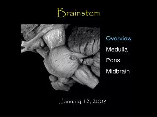
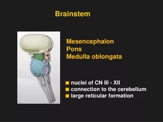
Current project:Imaging the brainstem at 1.5 and 4.0T • Why imaging the brainstem? • Optimizing sequence parameters: MORE anatomical information than the BEST known anatomy book* !!! * Henri M. Duvernoy’s: “Human Brain Stem Vessels”
Slice 3 7 5 3 5 1 4 3 1 4 2 2 8 6 6 1: Red nucleus vascularized part 2: Red nucleus non-vascularized part 3: Substantia nigra pars compacta 4: Substantia nigra pars reticulata 5: Crus cerebri 6: Lateral geniculate body 7: Fasicula nigrale 8: Capsule of Red nucleus 1
4T image quality in comparison to 1.5T image quality 4T magnitude image 4T phase image 1.5T magnitude image 1.5T phase image