Download
1 / 19
190 likes | 388 Views
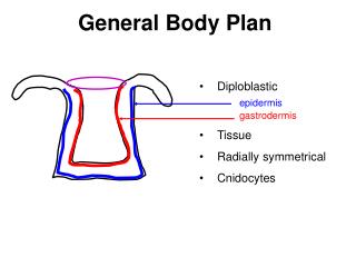
Comparative Anatomy General Body Plan. Note Set 2 Chapters 2 & 4 . Pharynx (fair-inks) with slits. Figure 3.1: Pharyngeal arches lateral view and (b) ventral cross section. Pharyngeal arches - associated with slits Anamniotes (lower vertebrates)- have gill slits

E N D
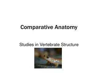
Comparative AnatomyGeneral Body Plan Note Set 2 Chapters 2 & 4
Pharynx (fair-inks) with slits • Figure 3.1: Pharyngeal arches • lateral view and • (b) ventral cross section. • Pharyngeal arches- associated with slits • Anamniotes (lower vertebrates)- have gill slits • Amniotes- have slits in embryo but gills never form • Endodermal pouches- pharyngeal pouch that grows toward surface of animal • Ectodermal groove- pharyngeal groove on outside that grows toward each pouch
Embryo Figure 3.2: Embryo pharyngeal arches. Figure 3.3: Embryo development at 1 month.
(a) • In tetrapods, 1st pharyngeal slit becomes auditory tube and middle ear cavity • Aortic arches housed by pharyngeal arch (b) Figure 3.4: Early pharyngeal devel.. of shark (a) early stage (b) later stage.
Figure 3.5: Fate of the pharyngeal arches. • 1st (mandibular) arch- mandibular and maxillary portions • 2nd (hyoid) arch • Other arches are numerically named • Most vertebrates have 6 pairs of arches
Figure 3.6: Pharyngeal arches. • Each pharyngeal arch contains a cartilage, artery, mesoderm component, and cranial nerve.
Body Plan • Head • Trunk • Tail Figure 3.7: Sagittal section of craniate embryo.
Head • Head • Cephalization- development of sense organs • Protective covering of dermal bone (not replacement bone) or cartilage Figure 3.8.
Trunk • Somites- muscle masses beside notochord; embryonic structures that turn into bone • Coelom- body cavity between gut and body wall, lined by peritoneum Figure 3.10. Figure 3.9: Coelom.
Trunk (cont.) • Other visceral organ cavities: • Heart- pericardium • Lungs- pleura • Abdomen- peritoneum Figure 3.12: Pericardium and pleural cavities. Figure 3.11: Pericardium cavities.
Tail • From body to end of the digestive tract • Somites and notochord • Innervations • Dorsal and ventral aorta • Food storage, defense, locomotion
Pituitary Development • Stomodeal ectoderm plate pushes inward and forms pocket (Rathke’s pouch) that leads to anterior lobe • This invagination meets the dienchephalon’s infundibular process which forms the posterior lobe Figure 3.13 Figure 3.14
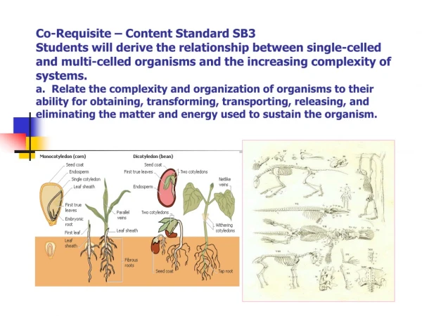
Characteristics of Vertebrates • Divided into two superclasses: Pisces and Tetrapoda • Demonstrate bilateral symmetry • Distinguishing features of vertebrates: • Presence of notochord • Pharynx with slits • Dorsal tubular nervous system • Developed vertebrae • Post anal tail
Notochord • Most primitive endoskeletal structure • Jawless fish- n.c. long and enlarged • Cartilagenous fish- n.c. surrounded by cartilagenous vertebrae • Bony fish and amphibians- n.c. surrounded by larger vertebrae • Amniotes- n.c. almost lost
Notochord (cont.) • In mammals, between successive centrum are disks • Within disks, the notochord is mostly replaced with pulpy nucleus Figure 3.15: Vertebrae. Figure 3.16: MRI scan of notochord.
More Vertebrate Features • Dorsal Nervous System • CNS- brain and spinal cord • Anamniotes- 10 pairs of cranial nerves • Amniotes- 12 pairs of cranial nerves • Vertebrae column • Backbones • Post-anal tail Figure 3.17: Vertebral column.
Anatomical Directions Figure 3.18. Figure 3.19.
Cranial Nerves • Olfactory • Optic • Oculomotor • Trochlear • Trigeminal • Abducens • Facial • Vestibulocochlear • Glossopharyngeal • Vagus Amniotes only: • Spinal Accessory • Hypoglossal Figure 3.20
Literature Cited Figure 3.1- http://www.nature.com/nrg/journal/v2/n11/images/nrg1101-858a-i1.gif Figure 3.2- http://www.ratbehavior.org/images/EmbryoPharyngealArches.jpg Figure 3.3- http://islam.org.hk/It_is_the_Truth/somites.htm Figure 3.4, 3.7, 3.8, 3.10, & 3.18- Kent, George C. and Robert K. Carr. Comparative Anatomy of the Vertebrates. 9th ed. McGraw-Hill, 2001. Figure 3.5- http://pharyngula.org/images/arch_fates.gif Figure 3.6- http://connection.lww.com/Products/sadler/imagebank.asp Figure 3.9- http://www.sci.nu.ac.th/biology/elearning/picture5/7_coelomate.jpg Figure 3.11- http://mywebpages.comcast.net/wnor/thoraxlesson4.htm Figure 3.12- http://faculty.southwest.tn.edu/rburkett/A&P%20body%20cavities.htm Figure 3.13- http://people.musc.edu/~wilburd/Head%20and%20Neck%20I/sld012.htm Figure 3.14- http://www.cushings-help.com/rathke.htm Figure 3.15- http://cal.vet.upenn.edu/saortho/chapter_62/62mast.htm Figure 3.16- http://www.telepathology.com/cases/forum/case17g.jpg Figure 3.17- http://anthro.palomar.edu/primate/footnote.htm Figure 3.19- http://encyclopedia.thefreedictionary.com/Terms%20for%20anatomical%20location Figure 3.20- http://www.besthealth.com/besthealth/bodyguide/reftext/html/nerv_sys_fin.html