Download
1 / 52
570 likes | 939 Views
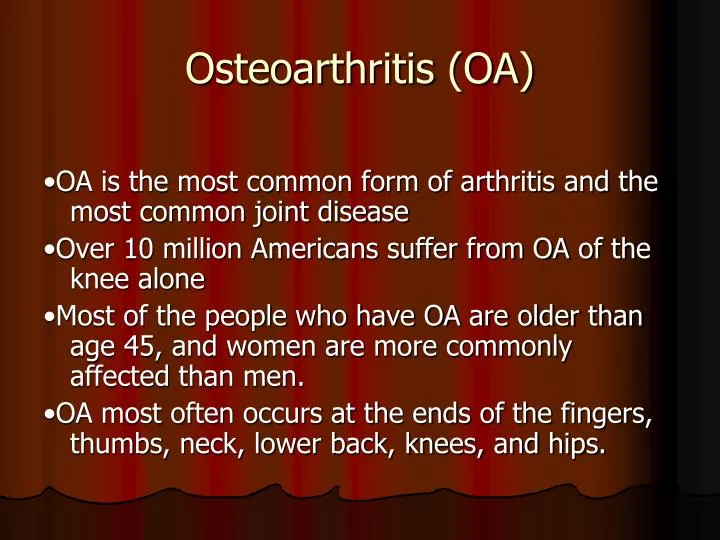
Osteoarthritis (OA). •OA is the most common form of arthritis and the most common joint disease •Over 10 million Americans suffer from OA of the knee alone •Most of the people who have OA are older than age 45, and women are more commonly affected than men.

E N D
Osteoarthritis (OA) •OA is the most common form of arthritis and the most common joint disease •Over 10 million Americans suffer from OA of the knee alone •Most of the people who have OA are older than age 45, and women are more commonly affected than men. •OA most often occurs at the ends of the fingers, thumbs, neck, lower back, knees, and hips.
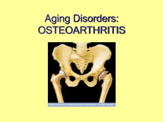
OA –Risk Factors AgeAge is the strongest risk factor for OA. Although OA can start in young adulthood, if you are over 45 years old, you are at higher risk.Female genderIn general, arthritis occurs more frequently in women than in men. Before age 45, OA occurs more frequently in men; after age 45, OA is more common in women. OA of the hand is particularly common among women. Joint alignmentPeople with joints that move or fit together incorrectly, such as bow legs, a dislocated hip are more likely to develop OA in those joints.
OA –Risk Factors Hereditary gene defect A defect in one of the genes responsible for the cartilage component collagen can cause deterioration of cartilage. injury Joint injury or overuse caused by physical labor or sports Traumatic injury (ex. Ligament or meniscaltears) to the knee or hip increases your risk for developing OA in these joints. Joints that are used repeatedly in certain jobs may be more likely to develop OA because of injury or overuse. Obesity Being overweight during midlife or the later years is among the strongest risk factors for OA of the knee.
OA –Symptoms • OA usually occurs slowly -It may be many years before the damage to the joint becomes noticeable • Only a third of people whose X-rays show OA report pain or other symptoms
OA –Symptoms • Steady or intermittent pain in a joint • Stiffness that tends to follow periods of inactivity, such as sleep or sitting • Swelling or tenderness in one or more joints [not necessarily occurring on both sides of the body at the same time] • Crunching feeling or sound of bone rubbing on bone (called crepitus) when the joint is used
Osteoarthritis (OA) -Definition Osteoarthritis may result from wear and tear on the joint•The normal cartilage lining is gradually worn away and the underlying bone is exposed. The repair mechanisms of tissue absorption and synthesis get out of balanceand result in osteophyte formation (bone spurs) and bone cysts
OA –Articular Cartilage Articularcartilageisthemaintissueaffected OA resultsin: • Increasedtissueswelling • Changeincolor • Cartilagefibrillation • Cartilageerosion down to subchondralbone
OA –Articular Cartilage Normal articular cartilage from 21-year old adult (3000X) Osteoarthritic cartilage (3000X)The surface changes alter the distribution of biomechanical forces further triggering active changes by the tissue
OA –Articular Cartilage • Thecartilagedamagecauseschondrocytecloningin an attempt to restorearticularsurface (Normaladultchondrocytesarefullydifferentiated and do not proliferate) • Normalarticularcartilage • Osteoarthriticcartilage
The newly dividing cells do not differentiate fully and cannot effectively synthesize the elements needed for matrix maintenanceThis results in a net loss of matrix components •Collagen content stays constant but fibrils are thinner and more disorganized -Decreased tensile strength •Proteoglycan loss results in an inability to hold on to water content:-Decreased resistance to compression –especially with repeated stress
OA –Overall Changes Osteoarthritis with lateral osteophyte, loss of articular cartilage and some subchondral bony sclerosis-X-ray shows loss of joint space
Osteoarthritis of theHand - diagnosis • Hand pain, aching, or stiffness and; • Hard tissue enlargement of two or more of 10 selected joints and; • Fewer than three swollen MCP (metacarpophalangeal) joints and; • Hard tissue enlargement of two or more DIP (distal interphalangeal) joints or deformity of two or more of 10 selected joints The 10 selected joints include: • Second and third DIP joints of both hands • Second and third PIP (proximal interphalangeal) joints of both hands • First CMC (carpometacarpal) joints of both hands
OA Osteoarthritis of theKnee - diagnosis • Kneepain and; At least three of the following 6 criteria: • 50 years of age or older • stiffness lasting less than 30 minutes, • crepitus, • bony tenderness, • bony enlargement, • no warmth to the touch
Osteoarthritis of theHip - diagnosis Hip pain and • Femoral and/or acetabularosteophytes evident on x-ray or • sedimentation rate less than or equal to 20 mm/hour and; • Joint space narrowing evident on x-rayAdditional criteria which are useful for diagnosing osteoarthritis of the hip: • Internal hip rotation of less than or equal to 15 degrees, • morning stiffness in the hip lasting less than or equal to 1 hour, and • age of 50 years or older are.
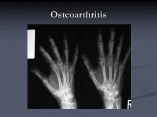
OA –Radiographic Diagnosis Asymmetrical joint space narrowingfrom loss of articular cartilage The medial (inside) part of the knee is most commonly affected by osteoarthritis.
OA –Radiographic Diagnosis •Asymmetrical joint space narrowing •Periarticularsclerosis •Osteophytes •Sub-chrondralbone cysts
Ostearthritic degenerated cartilage with exposed subchondral bone Normal Articular Cartilage OA –Arthroscopic Diagnosis Arthroscopy allows earlier diagnosis by demonstrating the more subtle cartilage changes that are not visible on x-ray
OA –Disease Management OA is a condition which progresses slowly over a period of many years and cannot be cured Treatment is directed at decreasing the symptoms slowing the progress of the condition Functional treatment goals: •Limit pain •Increase range of motion •Increase muscle strength
OA –Non-operative Treatments •Pain medications •Physical therapy •Walking aids •Shock absorption •Re-alignment through orthotics •Limit strain to affected areas
Proximal tibial osteotomy In the procedure to realign the angles, a wedge of bone is removed from the lateral side of the upper tibia. A staple or plate and screws are used to hold the bone in place until it heals. This converts the extremity from being bow-legged to knock-kneed.• The Proximal Tibial Osteotomy buys some time before ultimately needing to perform a total knee replacement. The operation probably lasts for 5-7 years if successful.