Download
1 / 54
570 likes | 973 Views
The Nervous System: The Spinal Cord & Spinal Nerves. Chapter 9 Anatomy & Physiology I. Outline. ROLE OF THE NERVOUS SYSTEM Structural divisions—anatomic (CNS and PNS) Functional divisions—physiologic (somatic and autonomic) NEURONS AND THEIR FUNCTIONS

E N D
The Nervous System: The Spinal Cord & Spinal Nerves Chapter 9 Anatomy & Physiology I
Outline • ROLE OF THE NERVOUS SYSTEM • Structural divisions—anatomic (CNS and PNS) • Functional divisions—physiologic (somatic and autonomic) • NEURONS AND THEIR FUNCTIONS • Structure of a neuron (cell body, cell fibers, myelin sheath) • Types of neurons (afferent, efferent, interneuron) • Nerves and tracts—bundles of neuron fibers • NEUROGLIA • Nonconducting cells • Protect and support nervous tissue • THE NERVOUS SYSTEM AT WORK • Nerve impulse (action potential) • Synapse—junction between neurons (neurotransmitter, presynaptic, postsynaptic, receptors) • SPINAL CORD • Location • Structure of the spinal cord • Reflex arc—pathway through the nervous system • Medical procedures involving the spinal cord • Diseases and other disorders of the spinal cord • SPINAL NERVES—31 PAIRS • Roots • Mixed nerves—combine sensory and motor fibers • Branches of the spinal nerves • Disorders of the spinal nerves—peripheral neuritis, sciatica, herpes zoster, Guillain-Barré • AUTONOMIC NERVOUS SYSTEM (VISCERAL NERVOUS SYSTEM) • Characteristics • Divisions of the autonomic nervous system • Cellular receptors • Functions of the autonomic nervous system
I. Role of the Nervous System • Chief coordinating center for all body systems • Conditions internally & externally are constantly changing. These changes stimuli. • Nervous system detects & responds to changes (stimuli) so the body can adapt & maintain homeostasis >> nerves carry message to and from Centers (Brain and the spinal cords) • Sensors- Nerves that detect stimuli • Effector – any tissue that carries out a command from the NS; they are muscles or glands
Anatomic Divisions of the Nervous System 1. Structural Division • Central nervous system(CNS) • Brain & • Spinal cord • Peripheral nervous system (PNS) • Cranial nerves • Spinal nerves • Nerves carrying messages to & from the body
2. Functional Division • Somatic nervous system • Controlled voluntarily • Effectors are skeletal muscles • No further subdivision • Autonomic nervous system • involuntarily controlled • Effectors: smooth muscle, cardiac muscle, glands • Subdivided into: • Sympathetic nervous system and • Parasympathetic nervous system
Neurons and Their Functions Neurons: Functional cell of the nervous system
Neuron Structure • Cell body: • Contains nucleus & organelles • Ganglion – a collection of nerve cell bodies that are located outside • Cell fibres: • Dendrite – fibers that conduct nerve impulses to the nerve body • Highly branched, tree-like appearance • Receptors of the NS – receive stimuli to NS • Axon - fibers that conduct nerve impulses away from the nerve body • Deliver impulses to another neuron, a muscle or a gland • Single long fiber
Myelin Sheath • Covering of fatty material over some axons that acts as an insulator • Schwann cell – a glial cell that produces myelin in the PNS in layers • Nodes of Ranvier – spaces between the myelin sheath • Nerve impulses jump from node to node, increasing their speed of conduction along an axon
Myelin Sheath - Neurilemma • Neurilemma – outer membrane of a Schwann cell that allows some peripheral nerves to repair themselves if injured • Neurilemma creates a sleeve into which a nerve can regenerate • CNS is not myelinated by Schwann cells so have no mechanism by which to repair themselves
White Matter vs Gray Matter • White matter – myelinated part of the NS • Gray matter – unmyelinatedpart of the NS
Types of Neurons • Sensory neurons (afferent neurons) • relay information to the CNS • Motor neurons (efferent neurons) • relay information from the CNS; efferent neurons • Interneurons • relay information within the CNS
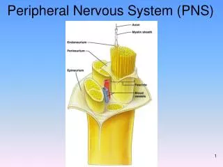
Nerves & Tracts • Nerve – a bundle of nerve fibers within the PNS • Tract – a bundle of nerve fibers within the CNS • Fascicles – a bundle of nerve fibers within a nerve or tract • Endoneurium – connective tissue surrounding an individual nerve fiber • Perineurium – connective tissue surrounding a neural fascicle • Epineurium – connective tissue surrounding an entire nerve
Neuroglia aka Glial Cells • Support nervous tissue & bind it to other structures (i.e. Schwann cells) • Aid in repair of cells • Act as phagocytes to remove pathogens & impurities (i.e. astrocytes) • Regulate the composition of fluids around cells • Can reproduce
The Nervous System at Work • The NS works by means of electrical impulses sent along neuron fibers and transmitted between cells at junctions
Nerve Impulse • Resting state a neuron at rest; polarized • Depolarization a neuron being stimulated, causing an electrical charge • Repolarization the electrical charge leaves and the neuron returns to its resting state
Nerve Impulse – Resting State • Resting state – a neuron at rest • Neurons at rest are polarized with a separation of + & - ions on either side of its plasma membrane (like a battery) • - ions are on the inside of the plasma membrane • Resting neuron has a negative charge • The separation of ions creates the potential for creating energy
Nerve Impulse - Depolarization • Depolarization – a neuron being stimulated, causing an electrical charge • An electrical, chemical or mechanical force stimulates the neuron • + ions (Na+ ions) rush into the neuron, causing the neuron to suddenly change from negative to positive • The nerve impulse starts
Nerve Impulse - Repolarization • Repolarization – the electrical charge leaves and the neuron returns to its resting state • The neuron cannot be stimulated until it returns to a negative state • The neuron must pump all of the + ions out; pumps out K+ • The Na+/K+ pump describes the action of the neuron pumping + ions Na+ & K+ back out of the cell • Requires energy (ATP)
Nerve Impulse • Action potential – the traveling of an electric charge along the plasma membrane of a neuron - < 1/1000 of a second
Synapse • Point of junction for transmitting a nerve impulse from one nerve axon to • Another nerve dendrite • A muscle at the neuromuscular junction • A gland • Synaptic cleft – the space between one cell & another • Information must cross this cleft for the nerve impulse to continue or create action • Can do this chemically, through neurotransmitters • Can also do this electrically, as in cardiac muscle, CNS, smooth muscle
Neurotransmitters • Chemical that crosses the synaptic cleft from the axon of one neuron to signal the receiving cell • Receptors – on the receiving dendrite, motor end plate or gland that respond to specific neurotransmitters
Neurotransmitters • Epinephrine aka adrenaline • ANS • Norepinephrine aka noradrenaline • ANS • Acetylcholine – • ANS • Acts between neurons & muscles at the neuromuscular junction
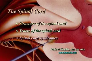
Spinal Cord • The link between the brain & the peripheral nervous system • Spinal cord ends around T12 and then a tail of spinal nerves called the cauda equina travels the rest of the way down the spinal canal
Spinal Cord Structure • Composed of both white (myelinated) & gray (unmyelinated) matter • White matter – outer layer • Gray matter – H shaped middle section • Dorsal horn – back part of H • Ventral horn – front part of H • Gray commissure – the middle bar of the H, connecting the dorsal & ventral horn Central canal: Contains cerebrospinal fluid Posterior median sulcus: divides the right and left portions of the posterior white matter Anterior median fissure : separates the right and left portions of the anterior white matter.
Spinal Cord Structure • Ascending tracts – sensory (afferent) impulses entering the spinal cord carrying information to the brain • Descending tracts – motor (efferent) impulses traveling from the brain to the body
Reflex Arc • The complete pathway through the nervous system from the stimulus to the body’s response • Five parts to the reflex arc 1. Receptor End of dendrite,receives stimulus 2. Sensory neuron afferent; carries messages to CNS from body 3. CNS processes the information & coordinates response 4. Motor neuron efferent; message sent from CNS to body 5. Effector a muscle or gland outside the CNS that carries out a response
Reflex Activities • Simple reflex – rapid automatic response • Involves very few neurons • a given stimulus always produces same response • Spinal reflex – reflex arc passes through the spinal cord & does not travel to the brain • Stretch reflex – muscle is stretched & responds by contracting • A simple spinal reflex
Spinal Nerves • 31 pair of nerves that attach directly to the spinal cord; all spinal nerves are mixed nerves • First part of the peripheral nervous system • Attach to spinal cord by 2 branches • Dorsal root – receives sensory information • Dorsal root ganglion – contains all sensory (afferent) neuron cell bodies • Ventral root – send motor information (efferent) to muscles and glands
Branches of the Spinal Nerves • Plexus – branches off the spinal nerves that intertwine into a network • Cervical plexus – supplies head & neck • phrenic nerve to diaphragm • Brachial plexus – shoulder and upper extremities • Lumbosacral plexus – pelvis & legs • Sciatic nerve – largest nerve in body; to legs
Dermatome • Dermatome the area of skin supplied by a single spinal nerve
Autonomic Nervous System • Regulates glands, smooth muscle & cardiac muscle • Automatic and without conscious awareness • Motor Pathway (efferent) different in ANS than in somatic NS (voluntary) because has 2 motor nerves • Spinal cord to ganglion via preganglionic neuron • Ganglion to effector via postganglionic neuron • Somatic NS has a single motor neuron that travels from spinal cord directly to effector
Divisions of the Autonomic NS • Sympathetic – “fight or flight” responses of organs • Accelerates organs that are needed for flight or fight • Brakes organs that are not needed for flight or fight • Parasympathetic • Brings organs back into balance after stress or danger has been alleviated • Reverses fight or flight effects • Most organs are innervated by both sympathetic & parasympathetic fibers
Sympathetic NS Structure • Thoracolumbar – preganglionic efferent motor fibers begin in spinal cord at around T1 and continue to about L2 • Synapse with sympathetic ganglia near the spinal cord • Sympathetic chain – close to spinal cord from lower neck to upper abdomen • Collateral ganglia – farther from spinal cord in lower abdomen & pelvis • Celiac ganglion – digestive organs • Superior mesenteric ganglion – small intestine • Inferior mesenteric ganglion – large intestine, urinary & reproductive organs • Postganglionic fibers continue from ganglia to effector organs & glands • Act on effector organs by releasing epinephrine & norepinephrine
Parasympathetic NS Structure • Craniosacral – preganglionic efferent motor fibers begin at the brainstem & then begin again at the sacrum • Synapse with ganglia near the effector organs • Terminal ganglia • Postganglionic efferent motor fibers from ganglia to effector organs • Act on effector organs by releasing acetylcholine