Download
1 / 45
700 likes | 2.59k Views
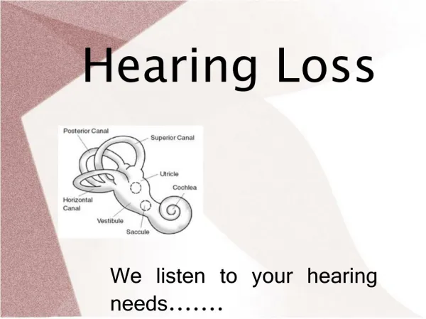
Genetic Hearing Loss. Jing Shen M.D. Ronald Deskin M.D. UTMB Dept of Otolaryngology March 2004. Epidemiology. Hearing loss occurs in 1 out of every 1,000 births 50 % are hereditary Syndromic vs. nonsyndromic 30% syndromic 70% nonsyndromic

E N D
Genetic Hearing Loss Jing Shen M.D. Ronald Deskin M.D. UTMB Dept of Otolaryngology March 2004
Epidemiology • Hearing loss occurs in 1 out of every 1,000 births • 50 % are hereditary • Syndromic vs. nonsyndromic • 30% syndromic • 70% nonsyndromic • Autosomal dominant vs. autosomal recessive vs. x-linked vs. mitochondrion
Methods • Linkage mapping • Mouse model • Difficulties: • Families too small for linkage analysis • Assortive mating introducing various genes into one single pedigree • Incomplete penetrance
Syndromic deafness • Has other abnormalities • About 20-30% of genetic hearing loss • Two syndromes can be caused by different mutations of the same gene • Mutations of more than one gene can cause the same clinical phenotype
Alport syndrome • At least 1% of congenital hearing loss • X-linked inheritance (80%), autosomal recessive as well as dominant • Sensorineural hearing loss: mostly affect high tone • Renal dysfunction • Microscopic hematuria • Man are more severely affected than woman • Onset in early childhood and progress to renal failure in adulthood • increased risk of developing anti-GBM nephritis after renal transplantation
Alport syndrome • Ocular abnormalities • Lenticulus • Retina flecks • Defective collagen type 4 causes abnormalities in the basement membrane • 3 genes: COL4A5, COL4A3, COL4A4 • These collagens found in the basilar membrane, parts of the spiral ligament, and stria vascularis • Exact mechanism of hearing loss is unknown
Branchio-oto-renal syndrome • 2% of profoundly deaf children • Autosomal dominant disorder • Otologic anomalies: • variable hearing loss (sensorineural, conductive or mixed) • malformed pinna, preauricular pits • Branchial derived abnormalities: cyst, cleft, fistula • Renal malformation: renal dysplasia with anomalies of the collecting system, renal agenesis • Sometimes with lacrimal duct abnormalities: aplasia, stenosis • EYA1 gene mutation – knockout-mice showed no ears and kidneys because apoptotic regression of the organ primordia
Jervell and Lange-Nielsen syndrome • Autosomal recessive • 0.25% of profound congenital hearing loss • Prolonged QT interval, sudden syncopal attacks • Severe to profound sensorineural hearing loss • 2 genes identified: • KVLQT1: expressed in the stria vascularis of mouse inner ear • KCNE1 • Both gene products form subunits of a potassium channel involved in endolymph homeostasis
Norrie syndrome • X-linked inheritance • Ocular symptoms with congenital blindness: pseudotumor of the retina, retinal hyperplasia, hypoplasia and necrosis of the inner layer of the retina, cataracts, phthisis bulbi • Progressive sensorineural hearing loss • Mental deficiency • Norrin gene: encodes a protein related to mucins
Pendred Syndrome • Most common form of syndromal deafness- 4-10 % • Autosomal recessive disorder • Sensorineural hearing loss • bilateral, severe to profound, and sloping in the higher frequencies • incomplete partition of the cochlear
Pendred syndrome • Vestibular dysfunction: • enlargement of the vestibular aqueducts, the endolymphatic sac and duct • Thyroid goiter: • usually euthyroid, can be hypothyroid • defective organic binding of iodine • positive potassium perchlorate discharge test
Pendred syndrome • PDS gene mutations: • on chromosome 7q31 • encodes pendrin: an anion transporter in inner ear, thyroid, kidney • PDS knockout mouse: • complete deaf • endolymph-containing spaces enlargement • inner and outer hair cell degeneration • no thyroid abnormality
Stickler syndrome • Autosomal dominant • Variable sensorineural hearing loss • Ocular symptoms: progressive myopia, resulting in retina detachment and blindness • Arthropathy: premature degenerative changes in various joints • Orofacial features: midface hypoplasia • Three genes: COL2A1, COL11A1, COL11A2 • Associated with defective collagen protein • Each gene mutation corresponding to a phenotype
Treacher-collins syndrome • Autosomal dominant with variable expression • Conductive hearing loss • Craniofacial abnormalities: • Coloboma of the lower lids, micrognathia, microtia, hypoplasia of zygomatic arches, macrostomia, slanting of the lateral canthi • TCOF1 gene: • Involved in nucleolar-cytoplasmic transport • mutation results in premature termination of the protein product
Usher syndrome • Autosomal recessive disorder • Sensorineural hearing loss • Progressive loss of sight due to retinitis pigmentosa • Three different clinical types • 11 loci and 6 genes have been identified
Usher syndrome • Type 1: • Profound congenital deafness, absent vestibular response, onset of retinitis pigmentosa in the first decade of life • Type 2: • Sloping congenital deafness, normal vestibular response, onset of retinitis pigmentosa in first or second decade of life • Type 3: • Progressive hearing loss, variable vestibular response, variable onset of retinitis pigmentosa
Usher syndrome • MYO7A: encodes for myosin 7A, molecular motor for hair cells • USH1C: encodes for harmonin, bundling protein in stereocilia • CDH23: encodes cadherin 23, an adhesion molecule may be important for crosslinking of stereocilia, also may be involved in maintaining the ionic composition of the endolymph • Myosin 7A, harmonin, and cadherin 23 form a transient functional complex in stereocilia
Waardenburg syndrome • About 2% of congenital hearing loss • Usually autosomal dominant • Dystonia canthorum • Pigmentary abnormalities of hair, iris and skin • Sensorineural hearing loss • 4 clinical subtypes
Waardenburg syndrome • Type 1: • With dystopia canthorum • Penetrance for hearing loss 36% to 58% • Wide confluent eyebrow, high broad nasal root, heterochromia irides, brilliant blue eyes, premature gray of hair, eyelashes, or eyebrows, white forelock, vestibular dysfunction • Type 2: • like type 1 but without dystopia canthorum • Hearing loss penetrance as high as 87%
Waardenburg syndrome • Type 3 (Klein-Waardenburg syndrome): • Type 1 clinical features + hypoplastic muscles and contractures of the upper limbs • Type 4 ( Shah-Waardenburg syndrome): • Type 2 clinical features + Hirschsprung’s disease • Five genes on five chromosomes have been identified
Waardenburg syndrome • Type 1 and type 3: • all associated with PAX3 gene mutation • Type 2: • Associated with dominant mutations of MITF gene • Associated with homozygous deletion of SLUG gene • MITF was found to activate the SLUG gene
Waardenburg syndrome • Type 4: • EDNRB gene – encodes endothelin-b receptor, development of two neural crest derived-cell lineages, epidermal melanocytes and enteric neurons • EDN3 gene – encodes endothelin-3, ligand for the endothelin-b receptor • SOX10 gene – encodes transcription factor
Non-syndromic deafness • About 70-80% of hereditary hearing loss • Autosomal dominant (15%): • 41 loci (DFNA) and 20 genes identified • Usually postlingual onset, progressive • Severity from moderate to severe • Majority of the hearing loss in middle, high or all frequencies • Autosomal recessive (80%): • 33 loci (DFNB) and 21 genes identified • Usually prelingual onset, non-progressive • Severity from severe to profound • All frequencies affected • X-linked (2-3%): • 4 loci (DFN) and 1 gene identified • Either high or all frequencies affected
Non-syndromic deafness • Identified genes encode: • Unconventional myosin and cytoskeleton proteins • Extracellular matrix proteins • Channel and gap junction components • Transcription factors • Proteins with unknown functions • More than one gene found in the same loci (DFNA2 and DFNA3) • Some genes cause autosomal dominant and autosomal recessive hearing loss • Some genes cause non-syndromic and syndromic hearing loss
Ion homeostasis • Potassium recycling to maintain high potassium concentration in endolymph • KCNQ4: encodes a potassium channel • SLC26A4: encodes an anion transporter, pendrin • 4 gap junction genes: GJB2, GJB3, DJB6, GJA1 • Encode connexin proteins • Function of gap junctions: molecular pores connecting two adjacent cells allowing small molecules and metabolites exchange
GJB2 (Gap Junction Beta 2) • The first non-syndromic sensorineural deafness gene to be discovered • On chromosome 13q11 • 50% of recessive non-syndromic hearing loss • Encodes connexin 26 • Expressed in stria vascularis, basement membrane, limbus, spiral prominence of cochlea • Recycling of potassium back to the endolymph after stimulation of the sensory hair cell • 80 recessive and 6 dominant mutations • 35delG mutation • One guanosine residue deletion from nucleotide position 35 • Results in protein truncation • High prevalence in Caucasian population • Screening test available
Transcription factors • POU3F4 • X-linked mixed hearing loss • Stapes fixation causing conductive hearing loss • Increased perilymphatic pressure • Causing the typical “gusher” during stapes footplate surgery – stapes-gusher syndrome • POU4F3 • Autosomal dominant hearing loss • Knockout mice fail to develop hair cells with subsequent loss of spiral and vestibular ganglia • EYA4 • TFCP2L3
Cytoskeleton proteins • Associated with actin-rich stereocilia of hair cells • Myosin: actin-dependent molecular motor proteins • MYH9 • MYO3A, MYO6, MYO7A, MYO15 – all have vestibular dysfunction • Otoferlin: calcium triggered synaptic vesicle trafficking • OTOF • one particular mutation accounts for 4.4% of recessive prelingual hearing loss negative for GJB2 mutation • Actin-polymerization protein: HDIA1 • Harmonin: organize multiprotein complexes in specific domains (tight junction, synaptic junction) • USH1C (also in Usher type 1c) • Cadherin: important for stereocilia organization • CDH23 ( also in Usher type 1d)
Extracellular matrix components • TECTA • Encodes alpha tectorin- component of the tectorial membrane • Knockout mice with detachment of tectorial membrane from the cochlear epithelium • COL11A2 • Encodes collage type XI polypeptide subunit 2 • Knockout mice with atypical and disorganized collagen fibrils of the tectorial membrane • COCH • Encodes COCH (coagulation factor C homologue) protein • Expressed in cochlear and vestibular organs • Associated with vestibular problems
Unknown function genes • WFS1 • Dominant sensorineural hearing loss • Responsible for 75% of low frequency nonsyndromic progressive hearing • Responsible for up to 90% of cases of Wolfram syndrome, a recessive disorder with diabetes mellitus, diabetes insipidus, optic atrophy, and deafness
Mitochondrial disorders • 2-10 mitochondrial chromosomes in each mitochondrion • Transmitted only through mothers • With syndromic hearing loss • Associated with systemic neuromuscular syndromes: such as Kearns-Sayre syndrome, MELAS, MERRF • Also in families with diabetes and sensorineural hearing loss • Associated with skin condition: palmoplantar keratoderma • With non-syndromic hearing loss • With aminoglycoside ototoxic hearing loss • A1555G mutation in the 12S ribosomal RNA gene • Maternally transmitted predisposition to aminoglycoside ototoxicity • Accounts for 15% of all aminoglycoside induced deafness
Evaluation • History • Prenatal: infection, medication • Perinatal: risk factors • Postnatal: infection, speech and language milestones • Family: • hearing loss in first and second degree relatives • Hearing loss occurred before age 30 • Consanguinity or common origin from ethnically isolated areas
Evaluation • Physical exam: features of syndromic hearing loss • Hair color: white forelock, premature graying • Facial shape • Skull shape • Eye: color, position, intercanthal distance, cataracts, retinal findings • Ear: preauricular pit, skin tags, shape and size of pinna, abnormality of EAC and TM • Oral cavity: cleft • Neck: brachial anomalies, thyroid enlargement • Skin: hyper/ hypopigmentation, café-au-lait spots • Digits: number, size, shape • Neurological exam: gait, balance
Evaluation • Audiologic evaluation • Lab testing: based on history and physical exam • Torch titers • CBC and electrolytes • Urinalysis • thyroid function test (perchlorate discharge test) • EKG • Radiological study: • CT temporal bone is the test of choice • Dilated vestibular aqueduct (>1.5mm at middle third or >2mm anywhere along its length) • Mondini malformation • Semicircular canal absence or dysplasia • Internal auditory canal narrowing or dilation • Renal ultrasound
Genetic screening • GJB2 • most common cause of severe to profound nonsyndromic recessive deafness • High prevalence of 35delG mutation • Small size of GJB2 gene • SLC26A4- most common cause of Mondini dysplasia or dilated vestibular aqueduct syndrome • EYA1- 30-40% of families with a branchio-oto-renal phenotype
Genetic counseling • Goal: • Cause of deafness • Other medical implication • Chance of recurrence in future children • Implications for other family members • Assist family in making choices that are appropriate for them • Team approach including clinical/medical geneticist, genetic counselor, social worker, psychologists • Consent need to be obtained for genetic testing
Cochlear gene therapy • Adenoid associated virus as vector • Routes of delivery • Safety concern • Hearing loss • Regional and distal dissemination
Resources for hereditary hearing loss • Hereditary hearing loss home page http://www.uia.ac.be/dnalab/hhh • Online Mendelian Inheritance in Man www.ncbi.nlm.nih.gov/Omim