Download
1 / 55
580 likes | 642 Views
Learn about anatomy, clinical findings, classifications, treatment options, operative and non-operative methods for managing forearm fractures effectively. Enhance your surgical knowledge and patient outcomes.

E N D
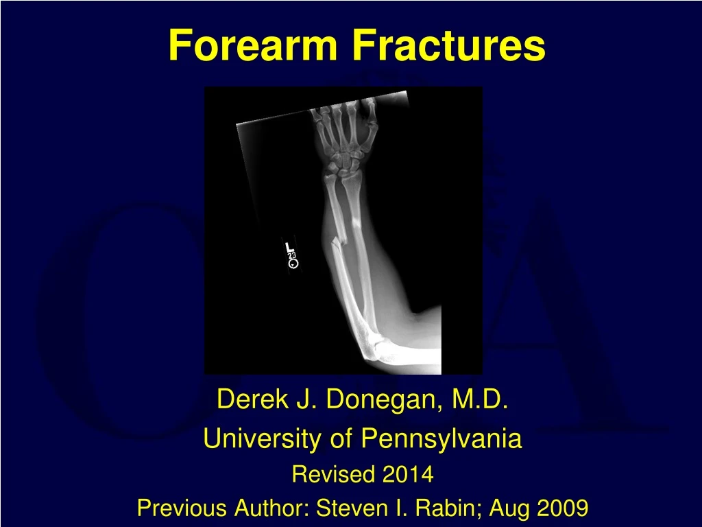
Forearm Fractures Derek J. Donegan, M.D. University of Pennsylvania Revised 2014 Previous Author: Steven I. Rabin; Aug 2009
Problem • Fractures of adult forearm are inherently unstable • According to the AO documentation center, forearm fractures accounted for 10-14% of all fractures between 1980 and 1996 • Mistreatment can lead to malunions and nonunions • Cosmetically unappealing • Functionally impeding
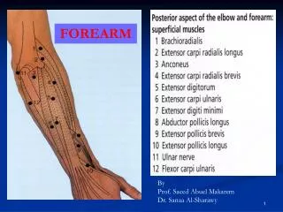
Anatomy • Radial Bow • Critical for rotation • Interosseous Membrane • Tethers Distal Ulna to Proximal Radius
Radial Nerve • PIN • Proximal Radial Neck • Superficial Branch Distal
Radial Artery • Posterior to Brachioradialis
Median Nerve • Midline • At risk with Carpal Tunnel • AIN along IOM
Mechanism • Low Energy • Direct blow (i.e. Nightstick fx) • Indirect • Galleazzi • Monteggia • High Energy • Associated injuries • open
Clinical Findings • PE • Floppy, Swelling, Pain • Assess Elbow and Wrist • Neurovascular Examination • AIN, PIN, radial/ulna arteries • Soft Tissue • Open Wounds • Compartments
Compartments • Dorsal: Extensors • Volar: Flexors • Superficial • Deep • Mobile Wad • BR • ECRB • ECRL
Compartment Syndrome • Pain • Passive Extension • High energy injury • Tx • Dorsal Approach • Volar Approach • Carpal Tunnel
Work-up • X-rays in 2 planes (AP and lateral) • Be sure to image joint above and below • Wrist and elbow • CT and MRI • Typically unnecessary • Add little clinical information
Classification • AO/OTA • 22 • Fracture type • A=simple • B=Wedge • C=complex • Involved bones • 1=ulna • 2=radius • 3=both bones
Type A • Simple Fracture • Ulna alone, Radius intact • Radius alone, Ulna intact • Both Bones broken • Ex: Transverse radius fracture
Type B • Wedge Fractures • Ulna alone • Radius alone • Both bones • Ex: Both Bones
Type C • Complex Fractures • Ulna alone • Radius alone • Both bones • Ex: both bones
Non-operative Poor Nonunion Malunion Non-operative Functional Brace / Cast Ulna Stable Closed Distal 1/3 < 10 Degrees Radius Nondisplaced Radial bow maintained Non-Operative Treatment
Operative Functional Anatomic All Unstable All Open Non-operative treatment rare Operative Treatment
Treatment • Early surgical intervention (within the first 6-8 hours) is optimal to avoid radioulnar synostosis • Goals • Anatomic reduction • Rigid fixation • Stable construct • Restoration of radial bow
Timing of Surgery • Early Surgery is Desirable but not Essential • Easier reduction especially if shortening • Avoids pre-op immobilization • Delayed Surgery • If poor soft tissues • If other injuries or medical problems prevent
Open Fractures • Antibiotics • Tetanus • Debridement • Irrigation • Surgical Tx • ORIF: Type I, II, IIIA • Ex-Fix: Type IIIB, IIIC
Treatment • Fixation options include • IM nailing • External fixation • plate fixation
Treatment • IM Fixation • Not routinely used • Soft tissue injury • Pathologic Fracture
Treatment • External Fixation • open type IIIb • open type IIIc
Plate Fixation provides stable strong anatomic fixation eliminates need for external casting allows early functional motion with union rates over 95%. Obtain anatomic reduction Restore ulna & radial length Prevents subluxation of either proximal or distal radioulnar joints Restore rotational alignment Restore radial bow Essential for rotational function of forearm Treatment
Approaches • Ulna • exposed along the subcutaneous border between the flexor and extensor carpi ulnaris • dorsal cutaneous branch of the ulnar nerve • ≈5 cm proximal to the wrist joint • identify and protect
Approaches • Radius • Two approaches • Henry • Volar • Good for middle to distal third fractures • Thompson • Dorsal • Good for proximal to middle third fractures
Approaches-Henry (volar) • incision begins 1 cm lateral to the biceps insertion • extends distally to the radial styloid • Interval between brachioradialis and FCR • Identify radial artery and superficial radial n. • Protect PIN proximally
Approaches-Thompson (dorsal) • Incision begins just anterior to the lateral epicondyle • Extends distally towards the ulnar side of Lister’s tubercle • interval is developed between the ECRB and the EDC, exposing the supinator muscle • Identify PIN • 1cm proximal to its distal edge of supinator
Intra-op Tips • Supine w/ hand table • Tourniquet • Approach simpler fx 1st • Reduce and provisionally fix • Approach other fx • Reduce and plate with LCDC or LCP in compression mode • Goal of 6 cortices above and below with 3 screws over 4 or more holes on each side • Check and modify reduction of other bone • Plate with LCDC or LCP in compression mode • Goal of 6 cortices above and below with3 screws over 4 holes on each side • Confirm reduction with c-arm • Irrigate and close ulna wound first • Irrigate and close radial wound • If unable to close, VAC and return in 3-5 days to close vs STSG
The Role of Bone Grafting • Bone Graft if there is Severe Bone Loss or the patient has an Open Fracture Severely Compromising Local Biology • If >1/3 cortical circumference is lost, consider bone grafting because interfragmentary compression becomes impossible • But the standard teaching that >30% comminution “requires” grafting has been challenged where newer biologic techniques are used. • Wright, RR, Schmeling, GJ, and Schwab, J.P. The necessity of acute bone grafting in diaphyseal forearm fractures: a retrospective review. J. Orthop Trauma 11:288-94, 1997.
Technical Tips for Plate Fixation of Forearm Fractures • Use Indirect Reduction Techniques Preserving Soft Tissue Attachments • Periosteal stripping must be minimized • Narrow retractors placed to avoid penetration of interosseous membrane • Close or Skin Graft Open Wounds within 3-5 days
Post-op • Sterile dressing and sugartong splint • Closely monitor compartments • Low threshold to split dressing • POD#1 • Initiate digital ROM • Delay Wrist/Elbow ROM 3-5 days • Prevents hematoma formation
Follow-up • Forearm rotation is initiated as the patient's comfort allows • Usually 1st or 2nd week post-op • RTC @ 2 weeks, 6 weeks, 12 weeks, and 4-6 months postoperatively • AP/lat X-rays each visit • Activity modification to ADL’s only until fracture healed • 8-12 weeks • progressively return to a normal lifestyle.
Complications • Refracture after plate removal • Symptomatic hardware • Nonunion • Malunion • Infection • Neurologic injury • Compartment syndrome • Radioulnarsynostosis
Two Years Bone Density Does Not Normalize for 21 months Rossen, JW et al, JBJS 1991:73B:65-7. 4 to 20% Refracture Risk Usually through original fracture or screw hole Large plate (4.5 mm DCP) Nonunion Infection & Nerve Injury Pain may persist after plate removal Post-removal 67% Residual Symptoms 9% Worse Weather Exercise Skin or Tendon Irritation Mih, AD et al, CORR 1994:299:256-8 Pain & Hardware Removal
Malunion • Loss of motion with >10◦ of angulation • 5◦ loss of radial row = 15◦ loss of sup/pro • Decreased grip strength occurs with loss of the radial bow • Schemitsch, EH & Richards RR JBJS 1992:74A:1068-78 • Tx: Osteotomy and Repair
Nonunion • Poor biomechanics • Poor Technique • Stable construct • Too few screws • Improper compression • Soft tissue management • Initial Fracture • Open Injury • Comminuted fracture • Tx • Revision Fixation • Bone Grafting • Segmental bone loss • Iliac crest <3.5cm • Consider vascularized fibular graft >3.5cm
Neurologic Injury • Closed Fracture • Usually Iatrogenic • PIN: Proximal approach • AIN: Vigorous Radial Reduction • Radial Sensory Branch: Anterior dorsal exposure • Open Fracture • AIN Most Common
Synostosis • Incidence 1-8% • Risks • BBFFx at same level • TBI • Surgical delay (> 2 wks) • Single incision • IOM Penetration • Tx • Early resection
Outcomes • Closed Fractures • 98% Union, 3% infection, 92% good function • Chapman, M et al: JBJS 1989:71A:159-69 • 96% Union, >85% good function • Anderson, LD et al: JBJS 1975:57A:287-97 • Open Fractures • 93% Union, 4% infection, 85% good function • Moed, BR et al: JBJS 1986:68A:1008-17
Outcomes • Motion • Near Normal • Grip Strength • 30% Reduced • Disability is Pain Related • Goldfarb et al JBJS Br 2005 Mar;87(3):374-9 • Droll et al JBJS Am 2007 Dec;89(12):2619-24
Special Cases • Fractures Associated with Joint Disruption • Galleazzi Fracture • Monteggia Fracture • Combined Patterns • Fractures Associated with other Injury • Floating Elbow (Ipsilateral Humerus Fracture) • Open Fractures
Fractures Associated with Joint DisruptionGaleazzi & Monteggia • Best Treatment • ORIF w. Plate Fixation of Diaphyseal Fracture • Joint Usually Reduces Indirectly and is stable • If Unstable: require open reduction of joint • If irreducible – it is usually because the diaphyseal fracture has been mal-reduced
Galeazzi Fractures • Classic: Fracture of distal 1/3 radial shaft with Dislocation Distal Radioulnar Joint • Variants: Fracture can occur anywhere along the radius or associated with fractures of both bones with DRUJ disruption
Galleazzi Fractures Radiographic Signs of DRUJ Injury: • Fracture at Base of Ulnar Styloid • Widened DRUJ on AP x-ray • Subluxed Ulna on Lateral x-ray • >5 mm Radial Shortening • Radius Fracture < 7.5cm from the wrist joint • (unstable DRUJ in 55%)
Galleazzi Fractures • Always require Plate fixation of the Radius • Distal Medullary canal too wide/funnel shaped for intramedullary fixation • Sometimes require temporary pin fixation of DRUJ or repair of the ulnar styloid when fractured • Postop: • If DRUJ stable – early motion • If DRUJ unstable – immobilize forearm in supination for 4-6 weeks in a long arm splint or cast • DRUJ pins are removed at 6-8 weeks
Galeazzi fractures • May be associated damage to triangular fibrocartilage, which may require early or late repair with open or arthroscopic techniques • Can Occur with Low Velocity Gunshots • Lenihan, MR et al J.O.T. 1992:6:32-35.
Monteggia FracturesClassic: Fracture of Proximal 1/3 Ulna with Dislocation of Radial Head
Monteggia Fractures Radiographic Findings: Normal: • Line Drawn through Radial Head & Shaft should always line up with Capitellum • Supinated Lateral: lines drawn tangential to head anteriorly and posteriorly should enclose the Capitellum Monteggia Fracture: These radiographic findings are disrupted
Monteggia Fractures • After fixation of the ulna, the radial head is usually stable (>90%) • If radial head not reduced recheck ulna length • If open reduction is required for the radial head, the annular ligament is repaired • Failure of the radial head to reduce with ulnar reduction is usually due to interposed annular ligament or rarely the radial nerve • Associated Radial Head Fractures may require fixation/replacement