Download
1 / 29
300 likes | 343 Views
Learn about the formation of the axial skeleton, including the skull, vertebrae, and ribs, through mesenchyme differentiation, endochondral ossification, and primary & secondary ossification centers. Understand spinal cord abnormalities like spina bifida.

E N D
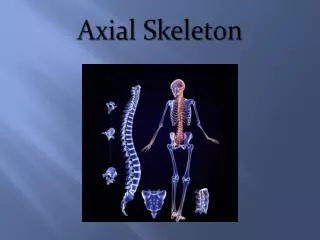
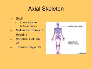
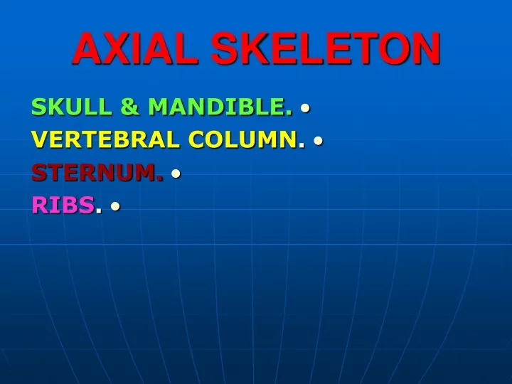
AXIAL SKELETON • SKULL & MANDIBLE. • VERTEBRAL COLUMN. • STERNUM. • RIBS.
PARAAXIAL MESODERM • It is longitudinal columns on each side of the notochord. • At the end of the 3rd week, it is differentiated into: • Somitomers (in the head region). • Somites (from the occipital region downward).
SOMITES • They are differentiated into : • Sclerotome (ventromedial). • It forms the vertebrae and ribs. • Dermomyotome(dorsolateral). • It forms the muscles and dermis of the skin.
MESENCHYME • At the end of the 4th week, the mesodermal cells form the polymorphous embryonic connective tissue (Mesenchyme). • The mesenchymal cells can migrate to different locations and are able to differentiate into : • Fibroblasts.Chondroblasts.Osteoblasts.
ENDOCHONDRAL OSSIFICATION • The mesenchymal cells first give rise to hyaline cartilage models, which in turn ossify.
MESENCHYMAL (Precartilagenous) Stage • In the 4th week, the mesenchymal cells from sclerotomes shift their position to be condensed around: • 1.Notochord • (around which the vertebrae develop).
MESENCHYMAL (Precartilagenous) Stage • 2.Neural tube(primordium of spinal cord). • 3. Body wall.
SCLEROTOME • Each sclerotome consists of: • Looselyarranged cells cranially. • Densely packed cells caudally.
SCLEROTOME • Some densely packed cells move cranially opposite the center of the myotome.
INTERVERTEBRAL DISC • The notochord degenerates in between the vertebrae. • It expands to form the Nucleus pulposus. • Later this nucleus is surrounded by theAnulus fibrosus(migrating densely packed cells). • These together constitute the intervertebral disc.
INTERVERTEBRAL DISC • Nerveslie in close relation to the inter vertebral discs.
CENTRUM • It is the primordiumofthe body of the vertebra. • It is formed from: • (1) the remaining densely packed cells which fuse with • (2) the loosely arranged of theimmediately caudal sclerotomes.
CENTRUM • It is an intersegmental structure. • Arterieslie on each side of the vertebral body. • The dorsal intersegmental arteries become the intercostal arteries.
VERTEBRAL (NEURAL) ARCH • It is formed from the mesenchymal cells around the neural tube.
THE VETEBRAL BODY • It is a composite of the anular epiphyses and the mass of bone between them. • It includes : • Centrum • Parts of the vertebral arch • Facets for the heads of the ribs.
THE CARTILAGENOUS (V. C.) • It begins in the 6thweek. • Two chondrification centers appear in each centrum . • They fuse with each other and with the centers of the vertebral arch.
THE CARTILAGENOUS (V. C.) • Spinousand Transverse processes: Formed from • extensions of Chondrification centers in the vertebral arch.
BONY VERTEBRAL COLUMN • Ossification begins during the embryonic period. • It endsat the age of 25years.
PRIMARY OSSIFICATION CENTERS • They are Threein number • One for the centrum (dorsal and ventral centers that fuse to form one) • One in each half of the vertebral arch. • Ossification is evident in the arch in the 8th week.
PRIMARY OSSIFICATION CENTERS • The bony halves of the neural arch fuse with each other in the first (3- 5) years. • The union starts first in the lumbar region then it progresses cranially.
NEUROCENTRALJOINTS • They are cartilagenous joints between the vertebral arch and the centrum. • They permit growth of the vertebral arches as the spinal cord expands. • These articulations disappear during the (3rd- 6th) years of age.
SECONDARYOSSIFICATIONCENTERS • They are five in number. • They appear afterpuberty. • They are : • One for the tip of the spinous process. • One for the tip of each transverse process. • Two Anular Epiphyses on the superior and inferior rami of the vertebral body.
SECONDARYOSSIFICATIONCENTERS • All secondary centers unite with the rest of the vertebra around the age of25 years. • Exceptions to typical ossification of vertebrae: • C1 (Atlas) • C2(Axis) • C7 • Lumbar,Sacrum and Coccyx.
SPINA BIFIDA • It is failure of fusion of the two halves of the vertebral arch.
SPINA BIFIDA • It occurs more frequently in girls than boys.
SPINA BIFIDA (OCCULTA) • It is a minor, insignificant anomaly of the vertebral column. It usually causes no clinical symptoms. • The skin over the bifid arch is intact • .
SPINA BIFIDA (OCCULTA) • There may be no external evidence of the vertebral defect. • Sometimes the anomaly is indicated by a tuft of hair. • The spinal cord and spinal nerves are usually normal.
SPINA BIFIDA (CYSTICA) • It is a severe type • The spinal cord and meninges are involved. • It is associated with neurologic symptoms.
RIBS • They are formed from mesenchymal costal processes of the thoracic vertebrae. • They are united with the vertebrae at the CostoVertebral joints. • They become cartilagenous and ossified before birth.