Download
1 / 50
500 likes | 774 Views
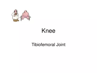
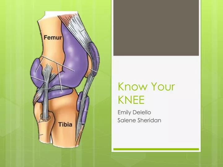
Know Your KNEE. Emily Delello Salene Sheridan . The knee joint is 3 joints in1. The Tibiofemoral joint is a hinge joint permitting flextion and extension. The Femoralpatellar joint is a plane joint where the patella glides across the distal end of the femur during knee flextion .

E N D
Know Your KNEE Emily Delello Salene Sheridan
The knee joint is 3 joints in1 • The Tibiofemoral joint is a hinge joint permitting flextion and extension. • The Femoralpatellar joint is a plane joint where the patella glides across the distal end of the femur during knee flextion. • Structurally it is a bicondylar joint allowing some rotation when the knee is partially flexed and when the knee is extending.
Patella Left and Right???
Rectus Femoris Origin: Anterior Inferior iliac spine Insertion : Tibial Tuberosity Action: Hip flexion, Knee extension Innervation: Femoral Nerve Vascular supply: Lateral circumflex artery
Vastusintermedialis Origin: Anterior femur Insertion: Tibial tuberosity via patellar tendon Action: Knee extension Innervation: Femoral nerve Vascular supply: Lateral circumflex femoral artery
Vastuslateralis Origin: Lateral aspera Insertion: Tibial tuberosity via patellar tendon Action: Knee extension Innervation: Femoral nerve Vascular supply: Lateral circumflex femoral artery
VastusMedialis Origin: Linea aspera Insertion: Tibial tuberosity via patellar tendon Action: Knee extension Innervation: Femoral nerve Vascular supply: Circumflex femoral artery
VastusLateralis VastusMedialis
Bicep Femoris Origin: Long head of the Ischial tuberosity. Short head the lateral lip of lineaaspera Insertion: Fibular head Action: Long head, extends hip and flexes knee. Short head, flexes knee Innervation: Long head, sciatic nerve. Short head, common peroneal nerve Vascular supply: Inferior gluteal artery
Semimembranous Origin: Ischial Tuberosity Insertion: Posterior surface of medial condyle of tibia Action: Extend hip and flex knee Innervation: Sciatic nerve Vascular supply: Inferior gluteal artery
Semimembranosus Semitendinosus
Semitendoninosus Origin: Ischial tuberosity Insertion: Anteromedial surface of Proximal tibia Action: Extend hip and flex knee Innervation: Sciatic nerve Vascular supply: Deep femoral
Popliteus Origin: Lateral condyle of femur Insertion: Posteriorly on medial condyle of tibia Action: Initiates knee flexion Innervation: Tibial nerve Vascular Supply: Popliteal artery
gastrocnemius Origin: Medial and lateral condyles of femur Insertion: Posterior calcaneus Action: Knee flextion, ankle plantar flextion Innervation: Tibial nerve Vascular supply: Popliteal artery
Lateral Head Medial Head of Gastrocnemius
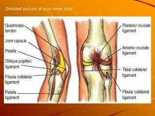
Ligaments A ligament is a tough band of fibrous tissue that connects bone to bone or bone to cartilage and supports and strengthens joints.
KEY • 1= Quadriceps femoris tendon • 2=Patellar Ligament • 3= Oblique popliteal ligament • 4=arcuate popliteal ligament • 5=Tibial collateral ligament • 6=Fibular collateral ligament • 7=Anterior cruciate ligament • 8=Posterior cruciate ligament • 9=Transverse ligament
1 2
4 4
7 5 6 3 8
1 2
Bursae-A bursa (plural bursae) is a small fluid-filled sac lined by synovial membrane with an inner capillary layer of viscous fluid (similar in consistency to that of a raw egg white). It provides a cushion between bones and tendons and/or muscles around a joint. This helps to reduce friction between the bones and allows free movement. Bursae are filled with synovial fluid and are found around most major joints of the body.
Bursae • 1=Prepatellar Bursa • 2=Deep Infrapatellar Bursa • 3=Suprapatellar Bursa • 4=Subcutaneous Infrapatellar Bursa
1 2 3