Download
1 / 68
690 likes | 831 Views
Practical Electrocardiography – Ventricular Tachycardia. Scott Ewing, D.O. Cardiology Fellow October 26, 2006. Terminology. Ventricular tachycardia (VT) – three or more ventricular extrasystoles in succession with rate > 120 bpm Non-sustained VT (NSVT) – lasts < 30 sec

E N D
Practical Electrocardiography –Ventricular Tachycardia Scott Ewing, D.O. Cardiology Fellow October 26, 2006
Terminology • Ventricular tachycardia (VT) – three or more ventricular extrasystoles in succession with rate > 120 bpm • Non-sustained VT (NSVT) – lasts < 30 sec • Sustained VT – lasts > 30 sec • Accelerated idioventricular rhythm – rate 100120 bpm
BBB Comparison • Normal leads V1 and V6 • Typical QRS-T patterns in RBBB and LBBB • Note the secondary T wave inversions in leads with an rSR' complex with RBBB and in leads with a wide R wave with LBBB
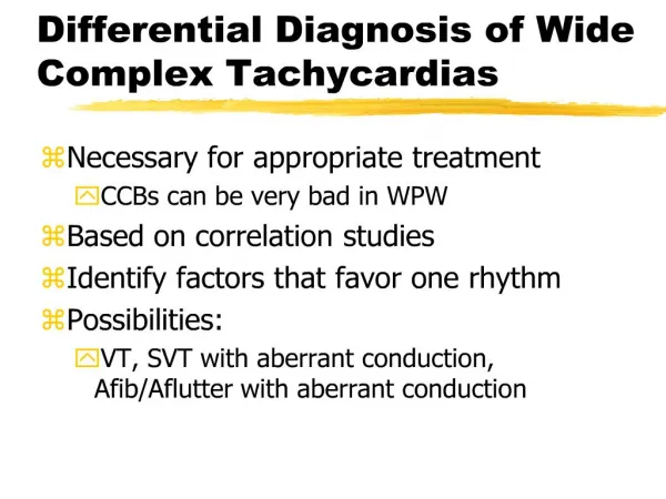
Varieties of Broad Complex Tachycardia • Ventricular - regular • Monomorphic ventricular tachycardia • Fascicular tachycardia • Right ventricular outflow tract tachycardia • Ventricular - irregular • Torsades de pointes tachycardia • Polymorphic ventricular tachycardia • Supraventricular • Bundle branch block with aberrant conduction • Atrial tachycardia with preexcitation
Etiology • VT usually a complication of severe heart disease • Early hours of MI, usually leading to ventricular fibrillation • Immediately after proximal obstruction of large artery • Chronically after large infarction, usually more stable form • May appear in “healthy” individuals • Originate from RV outflow tract • Posterior fascicle of the left bundle branch • Usually “cured” radiofrequency catheter ablation
Etiology • Many antiarrhythmic drugs proarrhythmic effects manifested as VT or torsades de pointes • Slow conduction and prolong the QRS complex and convert non-sustained VT into sustained VT • Prolong QTc interval and produce torsades de pointes • VT and proarrhythmic effect usually poor ventricular function and ischemic heart disease • Two aspects of the clinical history consistently predict a ventricular site of origin (90%) • Previous myocardial infarction • No previous tachyarrhythmia
Common Sources of Error • Believing VT cannot be well tolerated • Depending upon a single lead (Lead II) • Putting faith in irregularity • Depending upon independent atrial activity • Ignoring or neglecting QRS morphology
Believing That VT Cannot Be Well Tolerated • Commonly believed that VT is associated with a greater alteration of hemodynamics than are SVTs • Not true! Most are stable when first seen • Main factors that determine a patient's tolerance to a tachyarrhythmia of any origin • Ventricular rate • Size of the heart • Severity of underlying disease
Depending on a Single Lead • Lead II good for identifying P waves • Lead V1 (with its right-versus-left orientation) superior to lead II (with its base-to-apex orientation) for differentiating VT from an SVT • Lead V1 wide QRS complexes • V1-positive morphology, the differential diagnosis is between VT from the left ventricle and SVT with RBBB • V1-negative morphology, the differential diagnosis is between VT from the right ventricle and SVT with LBBB
Lead II vs. V1 • Left-ventricular tachycardia or SVT with RBBB vs. • Right-ventricular tachycardia or SVT with LBBB
Putting Faith in Irregularity • VT is sometimes irregular, so is SVT • When an intermittent irregularity appears in the morphology of the QRS complex, either on time or slightly early, the most likely cause is a breakthrough of conduction of the atrial rhythm to the ventricles • Fusion beat • Capture beat • If fusion or capture beats are proven, diagnosis is almost certainly VT • Usually at rates < 160 bpm • Not common
Putting Faith in Irregularity • 62 yo man who presented with acute SOB 2 months after an inferior–posterior MI • Arrows indicate capture beats and asterisks indicate fusion beats
Depending on Independent Atrial Activity • AV dissociation eliminates SVT originating from the atria or an accessory pathway, does not exclude SVT originating from AV node • Example of an AV-nodal tachycardia with LBBB aberrancy, confirmed by observing the similar QRS morphology occurring during sinus rhythm • However, this is the exception • With AV dissociation and wide-QRS-complex tachycardia, the diagnosis of VT is highly probable
AV Dissociation without VT • Recordings of limb leads I, II, and III during sinus rhythm (left) and AV-nodal tachycardia (right) • Arrows indicate the normally directed P waves visible in lead II without constant PR intervals
Ignoring or Neglecting QRS Morphology • Duration of wide QRS complexes may provide important diagnostic information, particularly if a recording is available of the patient's QRS-complex morphology during sinus rhythm • QRS width > 0.14 sec good indicator of VT • May also be seen in LBBB or RBBB • Less wide QRS (0.12 – 0.14 sec) not necessarily indicative of SVT with aberrancy • In LVT, V1-positive QRS complex usually described by • Monophasic R wave or • Diphasic qR complex, or occasionally • Triphasic RSR’ complex • When V1 QRS complex has two positive peaks (an R and an R’, with or without an S wave between these two peaks), termed “rabbit ears” • Relative heights of these “ears” have been used to differentiate LVT from SVT with right bundle-branch (RBBB) aberrancy • R > R’ suggests the presence of LVT because RBBB aberration is characterized by R’ > R
Ignoring or Neglecting QRS Morphology • 59 yo man after extensive anterior MI • Arrows taller first “rabbit ear” in lead V1 • Asterisks indicate the QS pattern in lead V6
Ignoring or Neglecting QRS Morphology • Absence of any positive deflection (QS or rS complexes) in V6 diagnostic for VT originating from either ventricle • These patterns could not be produced by RBBB or LBBB • Such patterns in lead V6 appear with LVT and RVT • Conversely, triphasic qRs morphology in V6, is virtually diagnostic of SVT with aberrancy
Ignoring or Neglecting QRS Morphology • 63 yo man with recurrent VT and heart failure awaiting cardiac transplantation while being maintained on propafenone therapy • Asterisks indicate the QS appearance in lead V6.
Ignoring or Neglecting QRS Morphology • Concordance precordial QRS complex directions another useful ECG clue • When all of the ventricular complexes from leads V1 to V6 are concordant negative or concordant positive • Diagnosis likely VT • Patterns highly atypical of either RBBB or LBBB • Concordant negativity virtually diagnostic of RVT • Concordant positivity virtually diagnostic of LVT
Ignoring or Neglecting QRS Morphology • Concordant negative (RVT) • Concordant positive (LVT)
Duration and Morphology of QRS Complex • Sequence of cardiac activation is altered, and the impulse no longer follows the normal intraventricular conduction pathway • Morphology of the QRS complex bizarre and duration prolonged (usually > 0.12 s) • Broader the QRS complex, more likely VT, esp. QRS > 01.6 s • QRS duration may exceed 0.2 s, particularly with electrolyte abnormalities, severe myocardial disease, or with antiarrhythmic drugs, such as flecainide • QRS complex in VT often has a RBBB or LBBB morphology • VT originating in the LV produces a RBBB pattern, whereas VT originating in the RV results in a LBBB pattern
Rate and Rhythm • VT rate normally 120300 bpm • Rhythm is regular or almost regular ( < 0.04 s beat to beat variation), unless disturbed by the presence of capture or fusion beats • If a monomorphic broad complex tachycardia has an obviously irregular rhythm the most likely diagnosis is atrial fibrillation with either aberrant conduction or preexcitation
Frontal Plane Axis • QRS axis normally - 30° to + 90°, with the axis most commonly ~60° • With VT mean frontal plane axis changes and often bizarre • Axis shift > 40° left or right suggestive of VT • Lead aVR is situated in the frontal plane at -210° • aVR negative with normal cardiac axis • aVR positive extremely abnormal axis to the left or right • aVR positive originates close to the apex, with depolarization moving upwards towards base of the heart
Direct Evidence of Independent Atrial Activity • With VT, sinus node continues to initiate atrial contraction • P waves are dissociated from the QRS complexes and are positive in leads I and II • Atrial rate usually slower than the ventricular rate • AV dissociation usually diagnostic for VT, lack of direct evidence of independent P wave activity does not exclude the diagnosis
Indirect Evidence of Independent Atrial Activity • Capture beat • Occasionally an atrial impulse may cause ventricular depolarization via the normal conduction system • The resulting QRS complex occurs earlier than expected and is narrow • Such a beat shows that even at rapid rates the conduction system is able to conduct normally, thus making a diagnosis of supraventricular tachycardia with aberrancy unlikely • Capture beats are uncommon, and though they confirm a diagnosis of ventricular tachycardia, their absence does not exclude the diagnosis
Indirect Evidence of Independent Atrial Activity • Fusion beats • Fusion beat occurs when a sinus beat conducts to the ventricles via the AV node and fuses with a beat arising in the ventricles • As the ventricles are depolarized partly by the impulse conducted through the HisPurkinje system and partly by the impulse arising in the ventricle, the resulting QRS complex has an appearance intermediate between a normal beat and a tachycardia beat • Fusion beats are uncommon, and though they support a diagnosis of ventricular tachycardia, their absence does not exclude the diagnosis
Indirect Evidence of Independent Atrial Activity • QRS concordance throughout the chest leads • QRS complexes in precordial leads are either predominantly positive or predominantly negative • Presence of concordance suggests that the tachycardia has a ventricular origin • Positive concordance probably indicates that the origin of the tachycardia lies on the posterior ventricular wall; the wave of depolarization moves towards all the chest leads and produces positive complexes • Similarly, negative concordance is thought to correlate with a tachycardia originating in the anterior ventricular wall
Right Ventricular Outflow Tract Tachycardia • Tachycardia originates from RVOT and impulse spreads inferiorly • Typically right axis deviation with a LBBB pattern • Tachycardia may be brief and self terminating or sustained • May be provoked by catecholamine release, sudden changes in heart rate, and exercise • Usually responds to β-blockers or CCB • Occasionally stops with adenosine treatment and so may be misdiagnosed as a supraventricular tachycardia
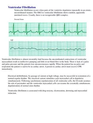
Torsades de pointes Tachycardia • Torsades de pointes (“twisting of points”) is a type of polymorphic ventricular tachycardia in which the cardiac axis rotates over a sequence of 520 beats, changing from one direction to another and back again • QRS amplitude varies similarly, such that the complexes appear to twist around the baseline • Usually associated with conditions that prolong QT interval • Usually not sustained • Recur unless the underlying cause is corrected • May be prolonged • May degenerate into ventricular fibrillation • Transient prolongation of the QT interval is often seen in the acute phase of myocardial infarction and may lead to torsades de pointes • Management different from the management of other VTs
Torsades de pointes • 62 yo woman on diuretics with syncope • Syncope recurred during this ECG recording • Serum potassium concentration was 2.3 mEq/L
Polymorphic Ventricular Tachycardia • Polymorphic ventricular tachycardia has the electrocardiographic characteristics of torsades de pointes but in sinus rhythm the QT interval is normal • Much less common than torsades de pointes • If sustained often leads to hemodynamic collapse • Occur in acute myocardial infarction and may deteriorate into ventricular fibrillation • Must be differentiated from atrial fibrillation with preexcitation, as both have the appearance of an irregular broad complex tachycardia with variable QRS morphology