Download
1 / 55
600 likes | 716 Views
Explore the classification, structure, and functions of sensory receptors including pain, temperature, and proprioception. Understand sensory adaptation and the mechanics of thermoreceptors and nociceptors.

E N D
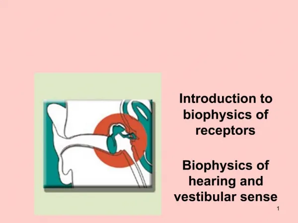
Marieb’s Human Anatomy and Physiology Marieb w Hoehn Chapter 13 – General Sensory Receptors Chapter 15 - Special Sensory Receptors Lecture 22
Lecture Overview • Introduction to the senses and sensation • Types of sensors • Classification of sensory receptors • Anatomy of the ear • Physiology of hearing/equilibrium • Anatomy of the eye • Physiology of vision Video 1 Video 2 Video 3
Background into Receptors… • What kinds of ‘messages’ does the brain understand? • What types of things in the environment do we have to respond to? • How do these environmental stimuli get converted into something the brain can understand? Electrical (nerve impulses) Light, sound, heat, cold, touch, etc. Receptors!
Receptors Classified by Location • Exteroceptors • - Respond to stimuli arising outside body • - Receptors in skin for touch, pressure, pain, and temperature • - Most special sense organs • Interoceptors (visceroceptors) • - Respond to stimuli arising in internal viscera and blood vessels • - Sensitive to chemical changes, tissue stretch, and temperature changes • - Sometimes cause discomfort but usually unaware of their workings
Classification by Receptor Structure • Simple receptors for general senses • Tactile sensations (touch, pressure, stretch, vibration), temperature, pain, and muscle sense • Modified dendritic endings of sensory neurons • Receptors for special senses • Vision, hearing, equilibrium, smell, and taste (Chapter 15)
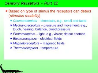
Sensory Receptors • Sensory Receptors • specialized cells or multicellular structures that collect information (transduce information into nerve impulses) • stimulate neurons to send impulses along sensory fibers to the brain (receptor vs. generator [action] potentials) • Chemoreceptors (general) • respond to changes in chemical concentrations • Pain receptors or nociceptors (general) • respond to stimuli likely to cause tissue damage • Thermoreceptors (general) • respond to changes in temperature • Mechanoreceptors (general, special) • respond to mechanical forces • Photoreceptors (special) • respond to light
Mechanoreceptors • Sense mechanical forces such as changes in pressure or movement of fluid • Two main groups • baroreceptors – sense changes in pressure (e.g., carotid artery, aorta, lungs, digestive & urinary systems) • proprioceptors – sense changes in muscles and tendons
Stretch Receptors - Proprioceptors • send information to CNS concerning lengths and tensions of muscles (and pressure, tension, and movement of joints) • 2 main kinds of proprioceptors • muscle spindles • in skeletal muscles • initiate contraction (mediates the stretch reflex) • Golgi tendon organs • in tendons • inhibit contraction
Stretch Receptors - Proprioceptors Muscle spindle – initiates contraction (stretch reflex) Golgi tendon organ – inhibits contraction
Sensory Adaptation • reduction in sensitivity of sensory receptors from continuous stimulation (painless, constant) • stronger stimulus required to activate receptors • smell and touch receptors undergo sensory adaptation • pain receptors usually do not undergo sensory adaptation (at level of receptor) • impulses can be re-triggered if the intensity of the stimulus changes
Temperature Sensors (Thermoreceptors) • Warm receptors • sensitive to temperatures above 25oC (77o F) • unresponsive to temperature above 45oC (113oF) • Cold receptors (3-4x more numerous than warm) • sensitive to temperature between 10oC (50oF) and 20oC (68oF) • unresponsive below 10oC (50oF) • Pain receptors are activated when a stimulus exceeds the capability (range) of a temperature receptor • respond to temperatures below 10oC • respond to temperatures above 45oC
Sense of Pain • pain receptors are callednociceptors • free nerve endings • Substance P or glutamate (inhib. by endorphins/enkephalins) • widely distributed • nervous tissue of brain lacks pain receptors (but meninges have nociceptors) • stimulated by tissue damage, chemical, mechanical forces, or extremes in temperature • nociceptors do not adapt (at the level of the receptor) • Visceral Pain • usually only type of visceral receptors that exhibit sensation • stretch, chemical irritation, ischemia (usu w/nausea) • may exhibit referred pain • not well localized
Special Senses • sensory receptors are within large, complex sensory organs in the head • hearing and equilibrium in ears • sight in eyes • smell in olfactory organs • taste (gustation) in taste buds
External Ear • auricle (pinna) • collects sounds waves • external auditory meatus • lined with ceruminous glands • carries sound to tympanic membrane • terminates at tympanic membrane • tympanic membrane • vibrates in response to sound waves
The Middle Ear (Tympanic Cavity) Typanic (attenuation) reflex: Elicited about 0.1 sec following loud noise; causes contraction of the tensor tympani m. and stapedius m. to dampen transmission of sound waves
Auditory Tube • Eustachian, auditory, or pharyngotympanic tube • connects middle ear to throat • helps maintain equal pressure on both sides of tympanic membrane • usually closed by valve-like flaps in throat When pressure in tympanic cavity is higher than in nasopharynx, tube opens automatically. But the converse is not true, and the tube must be forced open (swallowing, yawning, chewing).
Inner Ear • 3 Parts of Labyrinth • cochlea • functions in hearing • semicircular canals • function in equilibrium • vestibule • functions in equilibrium • utricle and saccule Labyrinth
Cochlea Cochlea as it would look ‘unwound’ • Scala vestibuli • upper compartment • leads from oval windowto apex of spiral • part of bony labyrinth • Scala tympani • lower compartment • extends from apex of the cochlea to round window • part of bony labyrinth
Organ of Corti • group of hearing receptor cells (hair cells) • on upper surface of basilar membrane • different frequencies of vibration move different parts of basilar membrane • particular sound frequencies cause hairs (stereocilia) of receptor cells to bend • nerve impulse generated
Physiology of Hearing Figure from: Marieb, Human Anatomy & Physiology, Pearson, 2013 Know pathway for exam Tympanic membrane malleus incus stapes oval window scala vestibuli scala tympani round window
Auditory Nerve Pathways Accessory Nerve (CN XI) Figure from: Martini, Fundamentals of Anatomy & Physiology, Pearson Education, 2004 (pons)
Vestibule • Utricle • communicates with saccule and membranous portion of semicircular canals • Saccule • communicates with cochlear duct • Macula • contains hair cells of utricle (horizontal) and saccule (vertical) Utricle and saccule provide sensations of: 1) gravity and 2) linear acceleration These organs function in static equilibrium (head/body are still)
Macula • responds to changes in head position • bending of hairs results in generation of nerve impulse
Semicircular Canals • three canals at right angles • ampulla (expansion) • swelling of membranous labyrinth that communicates with the vestibule • crista ampullaris • sensory organ of ampulla • hair cells and supporting cells • rapid turns of head or body stimulate hair cells Acceleration of fluid inside canals causes nerve impulse These organs function in dynamic equilibrium (head/body are in motion)
Crista Ampullaris Semicircular canals respond to rotational, nonlinear movements of the head
Pathways for Equilibrium Sensations For vestibulo-ocular reflex Figure from: Martini, Fundamentals of Anatomy & Physiology, Benjamin Cummings, 2004 *
External Anatomy of the Orbital Region Figure from: Saladin, Anatomy & Physiology, McGraw Hill, 2007
The Eye and Deep Orbital Region • Visual Accessory Organs • eyebrows • eyelids (palpebrae) • conjunctiva • lacrimal apparatus • extrinsic eye muscles Limbus
Eyelids • palpebrae = eyelids • composed of four layers • skin • muscle • connective tissue • conjunctiva • orbicularis oculi – closes eye (CN VII) • levator palpebrae superioris – raises eyelid (CN III) • tarsal (Meibomian) glands – secrete oil onto eyelashes; keep lids from sticking together • conjunctiva – mucous membrane; lines eyelid and covers portion of eyeball; keeps eye from drying out Fornix Sagittal section of right eye Figure from: Saladin, Anatomy & Physiology, McGraw Hill, 2007
Some External Disorders of Eye Sty(Infection of smaller glands (eyelashes) Chalazion(Infection of tarsal glands) Conjunctivitis(Inflammation of conjunctiva)
Lacrimal (Tear) Apparatus • lacrimal gland • lateral to eye • secretes tears • canaliculi • collect tears • lacrimal sac • collects from canaliculi • nasolacrimal duct • collects from lacrimal sac • empties tears into nasal cavity Tears: - supply oxygen and nutrients to cornea (avascular) - are antibacterial (contain antibodies and lysozyme) - lubricate and bathe the conjunctiva
Extraocular Eye Muscles • Superior rectus • rotates eye up and slightly medially • Inferior rectus • rotates eye down and slightly medially • Medial rectus • rotates eye medially
Extrinsic Eye Muscles • Lateral rectus • rotates eye laterally • Superior oblique • rolls eye, rotates eye down and laterally • Inferior oblique • rolls eye, rotates eye up and laterally Which cranial nerves innervate each of the muscles in the diagram above? LR6SO4AO3
Extraocular Eye Muscles & their CN Which cranial nerves innervate each of the muscles in the diagram above? LR6SO4AO3
Structure of the Eye - Overview Figure from: Martini, Fundamentals of Anatomy & Physiology, Pearson Education, 2004 Three layers (tunics) of the eye: - Outer fibrous tunic - Sclera and cornea - Middle vascular tunic (uvea) – Iris, ciliary body, and choroid - Inner neural tunic - Retina
Outer (Fibrous) Tunic • Cornea • anterior portion • transparent • light transmission • light refraction • well innervated • avascular • Sclera • posterior portion • opaque • protection • support • attachment site for extrinsic eye muscles Transverse section, superior view
Aqueous Humor • fluid in anterior cavity of eye • secreted by epithelium on inner surface of the ciliary processes • provides nutrients • maintains shape of anterior portion of eye • leaves cavity through canal of Schlemm (scleral venous sinus)
Lens • transparent, avascular • biconvex • lies behind iris • largely composed of lens fibers • enclosed by thin elastic capsule • held in place by suspensory ligaments of ciliary body • focuses visual image on retina (Crystallins) Loss of lens transparency = cataracts
SEM of Lens Figure from: Saladin, Anatomy & Physiology, McGraw Hill, 2007
Accommodation • changing of lens shape to view objects nearby Far vision (emmetropia)(20 ft. or greater) Presbyopia is the loss of the ability to accommodate with age Near vision
Middle (Vascular) Tunic = Uvea • 1. Iris • anterior portion • pigmented CT • controls light intensity • 2. Ciliary body • anterior portion • pigmented • holds lens • muscles reshape lens for focusing • aqueous humor • 3. Choroid coat • provides blood supply • pigments absorb extra light This layer contains the intrinsic muscles of the eye- Regulate the amount of light entering the eye - Regulate the shape of the lens
Iris • composed of connective tissue and smooth muscle • pupil is hole in iris • dim light stimulates (sympathetic) radial muscles and pupil dilates • bright light stimulates (parasympathetic, CN III) circular muscles and pupil constricts mydriasis miosis How would viewing near objects affect pupil size?
Inner (Neural) Tunic • retina • containsvisual receptors • continuous with optic nerve • ends just behind margin of the ciliary body • composed of several layers • macula lutea – yellowish spot in retina surrounds fovea • fovea centralis – center of macula lutea; produces sharpest vision; only cones • optic disc – blind spot; contains no visual receptors • vitreous humor – thick gel that holds retina flat against choroid coat Visual axis Transverse section, superior view
Optic Disc (Blind Spot) Figure from: Martini, Fundamentals of Anatomy & Physiology, Benjamin Cummings, 2004
Layers of Retina • receptor cells, bipolar cells, and ganglion cells - provide pathway for impulses triggered by photoreceptors to reach the optic nerve • horizontal cells and amacrine cells – modify impulses
Visual Receptors • Rods • long, thin projections • contain light sensitive pigment calledrhodopsin • hundred times more sensitive to light than cones • provide vision in dim light • produce colorless vision • produce outlines of object • view off-center at night • Cones • short, blunt projections • contain light sensitive pigments called erythrolabe, chlorolabe, and cyanolabe (photopsins) • provide vision in bright light • produce sharp images • produce color vision Dark adaptation by the rods takes approximately 30 minutes. This adaptation can be destroyed by white light in just milliseconds
Visual Pathway The right side of the brain receives input from the left half of the visual field The left side of the brain receives input from the right half of the visual field Figure from: Martini, Fundamentals of Anatomy & Physiology, Benjamin Cummings, 2004
Touch and Pressure Senses Class of mechanoreceptor