Download
1 / 38
470 likes | 859 Views
Pregnancy Diagnosis. Obstetrics and Gynecology Hospital of FudanUniversity Xing Chen, MD. Email: xing_chen2003@hotmail.com.

E N D
Pregnancy Diagnosis Obstetrics and Gynecology Hospital of FudanUniversity Xing Chen, MD. Email: xing_chen2003@hotmail.com
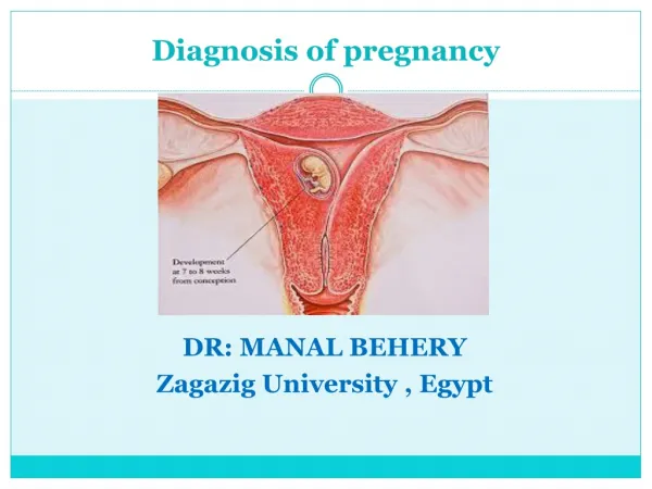
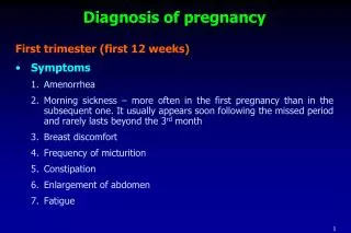
For a woman with regular menstrual cycles, a history of one or more missed periods following a time of sexual activity without effective contraception strongly suggests early pregnancy
Associated Symptoms • Fatigue • Nausea/vomiting • Breast tenderness
physical examination • softening and enlargement of the pregnant uterus • congestion and a bluish discoloration of the vagina (Chadwick sign) • softening of the cervix (Hegar sign) • Increased pigmentation of the skin • appearance of circumlinear striae on the abdominal wall (striae gravidarum) • Palpation of fetal parts • appreciation of fetal movement and fetal heart tones
human chorionic gonadotropin (hCG): α/β-subunit produced in the syncytiotrophoblast Urine approximately 4 weeks following the first day of the last menstrual period All urine pregnancy tests are best performed on early-morning urine specimens, which contain the highest concentration of hCG Serum specific and sensitive by following serial quantitative hCG levels and comparing them to the expected rise derived from normative data for proven normal intrauterine pregnancies Pregnancy test
Ultrasound examination • Abdominal ultrasound allowing visualization of a normal pregnancy gestational sac 5 to 6 weeks after the beginning of the last normal menstrual period (corresponding to β-hCG concentrations of 5000 to 6000 mIU/mL) • Transvaginal ultrasoundoften detects pregnancy at 3 to 4 weeks of gestation (corresponding to β-hCG concentrations of 1000 to 2000 mIU/mL)
Acoustic fetoscope beyond 18 to 20 weeks of gestational age Electronic Doppler devices approximately 12 weeks of gestation Detection of fetal heart activity “fetal heart tones”
Abnormal Pregnancy • Spontaneous abortion • Ectopic pregnancy • Trophoblastic disease
Prenatal diagnosisis the science of identifying structural or functional abnormalities-birth defects-in the fetus
Etiology of Birth Defects • Malformation • Deformation • Disruption • Other
Malformation • an intrinsic abnormality "programmed" in development, regardless of whether a precise genetic etiology is known • spina bifida
Deformation • caused when a genetically normal fetus develops abnormally because of mechanical forces imposed by the uterine environment • normal limb that develops contractures because of prolonged oligohydramnios
Disruption • which is a more severe change in form or function that occurs when genetically normal tissue is modified as the result of a specific insult • an amnionic band causing a cephalocele or limb-reduction abnormality
Other • Syndrome: trisomy 18 • Sequence: oligohydramnios leading to pulmonary hypoplasia • Association: VATER (association of vertebral defects, anal atresia, tracheoesophageal fistula with esophageal atresia, and radial dysplasia)
Techniques • Non-invasive • Minimally invasive • Invasive
Non-invasive techniques • Ultrasound • Magnetic Resonance Imaging (MRI)
Minimally Invasive Techniques • Cell free fetal DNA (cffDNA) • Pre-implantation genetic diagnosis (PGD)
Invasive Techniques • Chorionic villus sampling (CVS) • Amniocentesis • Percutaneous umbilical blood sampling (cordocentesis)
Key Guidelines • All women contemplating any form of prenatal diagnosis should be adequately counselled about the risks, benefits and limitations of any test, and provided with non-directional written information • Screening test for Down's syndrome and ‘20 week’ scan for structural anomalies • Women at risk of having a baby with congenital heart disease should be offered an extra fetal echocardiogram at 21–24 weeks • The middle cerebral artery Doppler peak systolic velocity can be used as a non-invasive method for diagnosing of fetal anaemia
Key Guidelines • Serial ultrasound measurements are of undoubted use in monitoring fetal growth but all formulae currently used to estimate fetal weight are inherently flawed and may give errors up to ±14% • MRI is a useful adjunct to ultrasound in prenatal diagnosis especially in the diagnosis of intra-cranial, intra-thoracic and gastrointestinal anomalies
Key Guidelines • Cell free fetal DNA testing has become widely established for the management of Rhesus disease and certain sex linked genetic disorders. With further research it is poised to offer much greater benefits in the field of minimally invasive prenatal diagnosis • Pre-implantation genetic diagnosis provides the opportunity for parents to avoid the distress of invasive testing and possible termination. However, the ethical and legal debate is set to continue for many years
Key Guidelines • CVS should not be performed before 10 weeks of gestation as it has been associated with limb reduction abnormalities. It appears to be safer if it is performed transabdominally rather than transcervically • Amniocentesis should not be performed at less than 15 weeks of gestation as before this it is associated with greater risk of pregnancy loss and possible talipes in the fetus
Key Guidelines • In experienced hands CVS and amniocentesis both carry a similar procedure related risk of miscarriage of 0.5–1% • Percutaneous umbilical blood sampling is now limited to potentially lifesaving in utero transfusion procedures for severe fetal anaemia
Neural-Tube Defects (NTDs) • anencephaly, spina bifida, cephalocele, and other rare spinal fusion (schisis) abnormalities • had higher levels of alpha-fetoprotein (AFP) in maternal serum and amnionic fluid
Maternal Serum AFP Screening influence factors: maternal weight, gestational age, diabetes, multifetal gestation
Evaluation of Maternal Serum AFP Elevation • genetic counseling • diagnostic test • Specialized Sonography • amniocentesis
Specialized Sonography • Transverse and sagittal images of the spine are increasingly used to characterize the size and location of spinal defects
Amniocentesis • amnionic fluid AFP level • assay for acetylcholinesterase
Down Syndrome • trisomy 18, 21 • First/second trimester: Sonography and maternal serum markers
Second-Trimester Screening • At 15 to 20 weeks • Triple test: • MSAFP (maternal serum alpha-fetoprotein ) • hCG or freeβ-hCG • uE3 (unconjugated estriol ) • Quadruple (Quad) test: + inh (dimeric inhibin alpha)
First-Trimester Screening • between 11 and 14 weeks • maternal serum analyte screening: • hCG (or free β-hCG) • pregnancy-associated plasma protein A (PAPP-A) • sonographic: nuchal translucency (NT) • combination of both
Be aware… • gestational age affects the accuracy • less sensitive in younger women
Be aware… • strong association between increasing nuchal translucency and fetal cardiac anomalies • nuchal translucency measurement is 3.5 mm or greater with a normal fetal karyotype, then targeted sonographic examination, fetal echocardiography, or both should be considered
Sonographic Screening for Aneuploidy • Major Structural Defects • "Soft Signs"
Diagnostic Techniques • Second-Trimester Amniocentesis between 15 and 20 weeks • Early Amniocentesis between 11 and 14 weeks • Chorionic Villus Sampling (CVS) at 10 to 13 weeks • Fetal Blood Sampling percutaneous umbilical blood sampling (PUBS) or cordocentesis • Fetal Tissue Biopsy • Preimplantation Genetic Diagnosis • Fetal Cells in the Maternal Circulation
Fetal Therapy -to improve the intrauterine environment • blood product transfusion • administration of medication transplacentally or via the fetal circulation • laser or radiofrequency ablation of vascular anastomoses • amnioreduction • shunt placement • fetal surgery
Reference • Obstetrics and Gynecology, 6th edition • Williams Obstetrics, 23rd edition • Prenatal diagnosis: Types and techniques. Early Human Development. 2012 (88) :3–8