Download

1 / 180
1.96k likes | 2.7k Views
Abdominal Examination. H.A.Soleimani MD Gastroenterologist. General principles of exam. Abdominal Examination. The History and Physical in Perspective. 70% of diagnoses can be made based on history alone. 90% of diagnoses can be made based on history and physical exam .

E N D
Abdominal Examination H.A.Soleimani MD Gastroenterologist
General principles of exam Abdominal Examination
The History and Physical in Perspective • 70% of diagnoses can be made based on history alone. • 90% of diagnoses can be made based on history and physical exam. • Expensive tests often confirm what is found during the history and physical.
Required Stethoscope Tongue blades Penlight Tape measure Sphygmomanometer Reflex hammer Safety pins Optional Gloves Gauze pads Lubricant gel Nasal speculum Turning fork: 128 Hz,512HzPocket visual acuity card Oto-ophthalmoscope Equipment for physical examination
Important aspects of physical examination----physician • Elegant appearance • Decent manner • Kind attitude • Highly responsibility • Good medical morals
Important aspects of physical examination---physician • Wash your hands, preferably while the patient is watching • Washing with soap and water is an effective way to reduce the transmission of disease
How to perform the physical examination? • Exposing only the area that are being examined • Offer a chaperone for both sexes. • Explain what you're going to do • Sequential
Important aspects of physical examination • The examiner should continue speaking to the patient • Showing care to his disease and answer to patient’s questions • It can not only release patient’s nerviness, but also help to establish the good physician-patient relationship
Gloves should be worn when.. • Examining any individual with exudative lesions or weeping dermatitis • When handling blood-soiled or body fluid-soiled sheets or clothing
General principles of exam • Good light • Relaxed patient • Full exposure of abdomen
General principles of exam • Have the patient empty their bladder before examination • Have the patient lie in a comfortable, flat, supine position • Have them keep their arms at their sides or folded on the chest
General principles of exam • Before the exam, ask the patient to identify painful areas so that you can examine those areas last • During the exam pay attention to their facial expression to assess for sign of discomfort
General principles of exam • Use warm hand, warm stethoscope, and have short finger nails • Approach the patient slowly and deliberately explaining what you will be doing
General principles of exam • Stand right side of the bed • Exam with right hand • Head just a little elevated • Ask the patient to keep the mouth partially open and breathe gently
General principles of exam • If muscles remain tense, patient may be asked to rest feet on table with hips and knees flexed
Other helpful points on examination • Take a spare bed sheet and drape it over their lower body such that it just covers the upper edge of their underwear
General principles of exam • If the patient is ticklish or frightened • Initially use the patients hand under yours as you palpate • When patient calms then use your hands to palpate. • Watch the patient’s face for discomfort.
Think Anatomically • When looking, listening, feeling and percussing imagine what organs live in the area that you are examining.
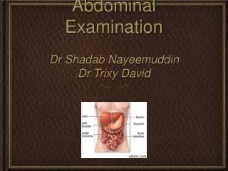
Right Upper Quadrant (RUQ) • liver, gallbladder, duodenum, right kidney and hepatic flexure of colon
Right Lower Quadrant (RLQ) • Cecum, appendix (in case of female, right ovary & tube)
Left Lower Quadrant (LLQ) • Sigmoid colon (in case of female, left ovary & tube)
Left Upper Quadrant (LUQ) • Stomach, spleen, left kidney, pancreas (tail), splenic flexure of colon
Epigastric Area • Stomach, pancreas (head and body), aorta
Landmarks of the abdominal wall, • Costal margin, umbilicus, iliac crest, anterior superior iliac spine, symphysis pubis, pubic tubercle, inguinal ligament, rectus abdominis muscle, xiphoid process.
Physical Examination of the Abdomen Inspection Auscultation Percussion Palpation Special Tests
Inspection Abdominal examination
Appearance of the abdomen • Is Aortic pulsation? • Is it flat or Scaphoid (Normally)? • Distended? • If enlarged, does this appear symmetric? • With bulging or moving?
Symmetrical in shape slightly full but not distended in older age group due to poor muscle tone or in subjects who are mildly overweight Scaphoid or flat in young patients of normal weight
Appreciation of abdominal contours • Standing at the foot of the table and looking up towards the patient's head. • Lower yourself until the anterior abdominal wall and ask the patient to breathe normally while you are doing so.
Appearance of the abdomen • Global abdominal enlargement is usually caused by air, fluid, or fat.
Appearance of the abdomen • Localized enlargement probably distend GB space occupying lesion, hepatomegaly….
An aortic aneurysm • Palpable mass • Patient feeling of pulsation • On rare occasions, a lump can be visible.
An aortic aneurysm • 1 in 10 men over 65 may have some enlargement of the abdominal aorta. About 1 in 100 will have a large aneurysm requiring surgery.
Appearance of the abdomen(Skin) • Abnormal venous patterns • Abnormal discoloration • Umbilicus issunken
Striae • Stretch marks are a light silver hue. • Pregnancy and obese individuals • Cushing’s syndrome (more purple or pink).
Appearance of the abdomen (Skin) • Tattoos • Scars can be drawn on schematic diagrams of the abdomen (a picture is worth a thousand words).
Cullen’s sign • Ecchymosis periumbilically. (intraperitoneal hemorrhage ruptured ectopic pregnancy, hemorrhagic pancreatitis..)
Grey-Turner’s sign • Ecchymosis of flanks. (retroperitoneal hemorrhage such as hemorrhagic pancreatitis)
Outward flow pattern from umbilicus in all directions ? Portal HTN
Evaluate venous return states • Place index finger side by side over a vein and press laterally, milking vein. • Release one finger and time refill, repeat with other finger. Venous return is in direction of faster filling.
Appearance of the abdomen • Areas which become more pronounced when the patient valsalvas are often associated with ventral hernias
More conspicuous in the thin than in the fat Greater in the old than in the young. Increased in thyrotoxicosis, hypertension, or aortic regurgitation) In those with an aortic aneurysm and tortuous aorta In those who have a mass joining the aorta to the anterior abdominal wall. Visible Pulsations
Gastric peristalsis is commonly seen in neonates with congenital hypertrophic pyloric stenosis Intestinal peristalsis in partial and chronic intestinal obstruction Colonic obstruction is usually not manifest as visible peristalsis Visible gastric Peristalsis Visible intestinal Peristalsis
Appearance of the abdomen Patient's movement • Patients with kidney stones will frequently writhe on the examination table, unable to find acomfortableposition