Download
1 / 23
240 likes | 464 Views
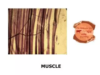
Muscle. 2 / 20. Movement with muscles. movement is one of the most prominent characteristics of animal life it can be either amoeboid , or more complicated using flagella , cilia or muscles

E N D
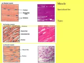
2/20 Movement with muscles • movement is one of the most prominent characteristics of animal life • it can be either amoeboid, or more complicated using flagella, cilia or muscles • Galenus (2.c. BC) – “animal spirit” is flowing from the nerves into the muscles causing swelling and shortening • spiral shortening of proteins was the supposed mechanism until the 50’s • new research techniques such as EM helped to elucidate the exact mechanism • muscles can be either smooth or striated • two subtypes of striated muscles are skeletal and heart muscle • mechanism of contraction is identical in all muscle types
3/20 Structure of the skeletal muscle Eckert: Animal Physiology, W.H.Freeman and Co., N.Y.,2000, Fig. 10-1.
4/20 Ultrastructure of the striated muscle Eckert: Animal Physiology, W.H.Freeman and Co., N.Y.,2000, Fig. 10-2.
5/20 Sarcomeres in cross-section Eckert: Animal Physiology, W.H.Freeman and Co., N.Y.,2000, Fig. 10-3.
6/20 Structure of the thin filament • G-actin: globular, 5.5 nm spheres • polymerized to “necklace” – two necklaces form a helical structure – F-actin • F-actins (length about 1000 nm, width 8 nm) are anchored to z-discs (-actinin) • in the groove of the F-actin tropomyosin (40 nm) troponin complexes are found • tropomyosin-troponin regulates actin-myosin interaction Eckert: Animal Physiology, W.H.Freeman and Co., N.Y.,2000, Fig. 10-5.
7/20 The thick filament • the thick filament is built up of myosin molecules • myosin molecules consist of two heavy chains (length 150 nm, width 2 nm) and 3-4 (species dependent) light chains • heavy chains form -helices twisted around each other bearing globular heads at the end • myosin molecules associate to form the thick filament (length 1600 nm, width 12 nm) • head regions are arranged into “crowns” of three heads at intervals of 14.3 nm along the thick filament • successive crowns are rotated by 40° resulting in a thick filament with 9 rows of heads along its length
8/20 Structure of the myosin filament Eckert: Animal Physiology, W.H.Freeman and Co., N.Y.,2000, Fig. 10-4, 6.
9/20 Sliding filament theory • during contraction A-band is unchanged, I-band shortens • length of actin and myosin filaments is unchanged • H.E. Huxley and A.F. Huxley independently described the sliding filament theory: actin and myosin are moving along each other • best proof is the length-tension curve, longer overlap stronger contraction • sliding is caused by the movement of cross-bridges connecting filaments • contraction is initiated by Ca++ ions released from the SR • excitation propagating on the sarcolemma is conducted to the SR by T-tubules invaginating at the level of z-disks Eckert: Animal Physiology, W.H.Freeman and Co., N.Y.,2000, Fig. 10-8.
10/20 Tubules in the muscle fiber Eckert: Animal Physiology, W.H.Freeman and Co., N.Y.,2000, Fig. 10-21.
11/20 Connection of T-tubules and SR Eckert: Animal Physiology, W.H.Freeman and Co., N.Y.,2000, Fig. 10-25.
12/20 Release of Ca++ ions • AP – spreads from the sarcolemma to the T-tubule – conformational change of the voltage-dependent dihydropyridin receptor – displacement or conformational change of the ryanodin receptor – Ca++ release • half of the ryanodin receptors are freeand are opened by the Ca++ ions - trigger Ca++ • restoration by Ca++-pump Eckert: Animal Physiology, W.H.Freeman and Co., N.Y.,2000, Fig. 10-4.
13/20 Mechanism of sliding • released Ca++ binds to the troponin complex, myosin binding site on actin is freed • cross-bridge cycle runs until Ca++ level is high • one cycle 10 nm displacement Eckert: Animal Physiology, W.H.Freeman and Co., N.Y.,2000, Fig. 10-16. Eckert: Animal Physiology, W.H.Freeman and Co., N.Y.,2000, Fig. 10-11.
14/20 Energetics of the contraction Eckert: Animal Physiology, W.H.Freeman and Co., N.Y.,2000, Fig. 10-29.
15/20 Types of muscle fibers • tonic fibers • postural muscles in amphibians, reptiles and birds • muscle spindles and extraocular muscles in mammals • no AP, motor axon forms repeated synapses • slow shortening – effective isometric contraction • slow-twitch (type I) fibers • mammalian postural muscles • slow shortening, slow fatigue – high myoglobin content, large number of mitochondria, rich blood supply – red muscle • fast-twitch oxidative (type IIa) fibers • specialized for rapid, repetitive movements – flight muscles of migratory birds • many mitochondria, relatively resistant to fatigue • fast-twitch glycolytic (type IIb) fibers • very fast contraction, quick fatigue • few mitochondria, relies on glycolysis • breast muscles of domestic fowl – white muscle
16/20 Motor unit • skeletal muscles in vertebrates are innervated by spinal or brainstem motoneurons – “final common pathway” • one fiber is innervated by only one motoneuron • one motoneuron might innervate several fibers (usually about 100) – motor unit • 1:1 synaptic transmission - 1 AP, 1 twitch • regulation of tension • AP frequency - tetanic contraction • recruitment – involvement of additional motor units • depending on the task, different types of fibers are activated – one motor unit always consists of fibers of the same type • type of muscle fibers can change, it depends on the innervation and the use – swapping of axons, change in type; difference between the muscles of a heavyweight lifter and a basketball player
17/20 Heart muscle • many differences, many similarities compared to skeletal muscles • pacemaker properties – myogenic generation of excitation • diffuse, modulatory innervation • individual cells with one nucleus • electrical synapses - functional syncytium • AP has plateau, long refractory period • voltage-dependent L-type Ca++-channels on T-tubules - entering Ca++ triggers Ca++ release from SR • Ca++ elimination: Ca++-pump (SR), Na+/Ca++ antiporter (cell membrane) - digitalis: inhibition of the Na/K pump - hypopolarization and increased Ca++ level • -adrenoceptor: IP3 - Ca++ release from SR • -adrenoceptor: cAMP - Ca++ influx through the membrane
18/20 Structure of the heart muscle Eckert: Animal Physiology, W.H.Freeman and Co., N.Y.,2000, Fig. 10-50.
19/20 Smooth muscle I. • not striated • actin filaments are anchored to the plasma membrane or to the dense bodies in the plasma • myosin filaments in parallel • single-unit smooth muscle • myogenic contraction • electrical synapses – synchronous contraction • contracts when stretched - basal myogenic tone • innervation modulates a few cells only through varicosities • in the wall of internal organs (gut, uterus, bladder, etc.) • multi-unit smooth muscle • neurogenic contraction • individual cells innervated by individual varicosities • e.g. pupil, blood vessels
20/20 Smooth muscle II. • activation by pacemaker cells, hormones, mediators released from varicosities • no fast Na+-channel • AP is not necessarily generated; it might have plateau if present • contraction is initiated by the increased level of Ca++ ions • Ca++influx through voltage/ligand-dependent channels, release from the SR (less developed) • instead of troponin-tropomyosin, caldesmon blocks the myosin binding site on actin – freed by Ca-calmodulin, or phosphorylation (PKC) • phosphorylation of myosin light chain (LC-kinase – activated by Ca-calmodulin) also induces contraction • light chain phosphorylation at another site by PKC - relaxation
Length-tension relation Eckert: Animal Physiology, W.H.Freeman and Co., N.Y.,2000, Fig. 10-8.
Role of the troponin complex Eckert: Animal Physiology, W.H.Freeman and Co., N.Y.,2000, Fig. 10-16.