Download

1 / 21
580 likes | 1.65k Views
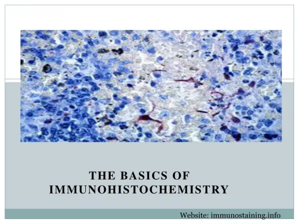
The basics of immunohistochemistry. Principle. Anigen (protein of interest) Primary antibody Secondary antibody. Immunohistochemistry – what’s good about it?. Antibodies bind to antigen in specific manner Can be used to locate particular cells and proteins

E N D
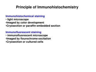
Principle Anigen (protein of interest) Primary antibody Secondary antibody
Immunohistochemistry – what’s good about it? • Antibodies bind to antigen in specific manner • Can be used to locate particular cells and proteins • Can be used to identify cellular events – e.g.apoptosis
Introduction • IHC takes its name from the roots • "immuno," in reference to antibodies used in the procedure, • and "histo," meaning tissue (compare to immunocytochemistry). • Immunohistochemistry is the localization of antigens or proteins in tissue sections • by the use of labeled antibodies as specific reagents • through antigen-antibody interactions • visualized by a marker such as fluorescent dye, enzyme, or colloidal gold
TYPES OF DETECTION • Visualising an antibody-antigen interaction can be accomplished in a number of ways. • Enzymatic staining • an antibody is conjugated to an enzyme, such as peroxidase, that can catalyse a colour-producing reaction • immunofluorescence • Alternatively, the antibody can also be tagged to a fluorophore, such as fluorescein or rhodamine
What cellular antigens can we target? • Cytoplasmic • Nuclear • Cell membrane
APPLICATIONS • disease diagnosis • drug development • and biological research
Types of IHC • Direct • Indirect
Direct method-primary antibody only • one step staining method • involves a labeled antibody reacting directly with the antigen in tissue sections. • utilizes only one antibody • procedure is short and quick. • However, it is insensitive due to little signal amplification and rarely used since the introduction of indirect method. anti-actinlabeled with 594
Indirect method – primary and secondary antibodies • involves an unlabeled primary antibody (first layer) which react with tissue antigen, • and a labeled secondary antibody (second layer) react with primary antibody • This method is more sensitive due to signal amplification • economic Donkey anti-goat labeled with 488 Goat anti-actin
IMP!! • Primary antibody • Should be raised against the antigen of interest • E.g. • for detecting human antigens- raise antibodies in specie other than humans! • HOW? • Injecting human MYOSIN protein in an animal specie e.g rabbit • Antibodies will be raised against human antigen in rabbit • These will be called as RABBIT ANTI-HUMAN MYOSIN ANTIBODY
Secondary antibody • Detects the Fc portion of the primary • must be against the antibody of the animal species in which the primary antibody has been raised) • E.g. • In our example primary antibody was rasied in – rabbit • Secondary antibody should detect this rabbit made antibody • So it must be an ANTI-RABBIT • Must be raised in another specie (other than humans & rabbits)
Sample preparation (FFPE) formalin fixed paraffin embedded • Tissue fixation • To ensure the preservation of tissue architecture and cell morphology, prompt and adequate fixation is essential • the most common fixative is formaldehyde (FF) • Embedding • in paraffin • to maintain the natural shape and • architecture of the sample during long-term storage and sectioning for IHC (PE) • Sectioning • Into slices as thin as 4-5 μm • with a microtome • Mounting • onto glass slides that are coated with an adhesive • 3-aminopropyltriethoxysilane (APTS) or poly-L-lysine • gelatin, egg albumin or Elmer's glue.
Antigen retrieval • What? • Retrieve your antigen for detection by IHC • Why? • Formaldehyde fixation generates methylene bridges • that crosslink proteins in tissue samples; • these bridges can mask antigen presentation and prevent antibody binding. • How? • to unmask the antibody epitopes, • either by heat (heat-induced epitope retrieval; HIER) • or enzymatic degradation (proteolytic-induced epitope retrieval; PIER).
Blocking Endogenous target activtiy • What? • Quenching or masking endogenous forms of enzymatic proteins (biotin, peroxidases or phosphatases) • Why? • When using Enzymatic detection • To prevent false positive and high background detection. • How? • Hydrogen peroxide – peroxidases • levamisole - Alkaline phosphatase
Blocking non-specific sites • What? • Masking sites that are similar to target sites • Why? • antibodies may partially or weakly bind to sites on nonspecific proteins that are similar to target • nonspecific binding causes high background staining that can mask the detection of the target antigen. • How? • Commonly blocking buffers are used • normal serum, non-fat dry milk, BSA or gelatin
Non-specific staining Before block After block
Controls • Postive control • to test a protocol or procedure and make sure it works. • It will be ideal to use the tissue that has the expression of your antigen • If the positive control tissue showed negative staining, the protocol or procedure needs to be checked until a good positive staining is obtained. • Negative control • To test for the specificity of an antibody involved • Exclude the primary antibody – no color should be obtained