Download

1 / 6
60 likes | 218 Views
Necrotising fasciitis . Photograph showing both groin wounds (day-12) with patchy areas of necrotic tissue and slough. Photograph showing both groin wounds (day-47) healing satisfactorily with evidence of a sinus in the upper part of the left groin wound (black arrow).

E N D
Photograph showing both groin wounds (day-12) with patchy areas of necrotic tissue and slough Photograph showing both groin wounds (day-47) healing satisfactorily with evidence of a sinus in the upper part of the left groin wound (black arrow) http://www.springerimages.com/Images/MedicineAndPublicHealth/5-10.1186_1477-7819-6-67-3
Photograph showing both groin wounds and perineum (day-20) with VAC system in place http://www.springerimages.com/Images/MedicineAndPublicHealth/5-10.1186_1477-7819-6-67-3
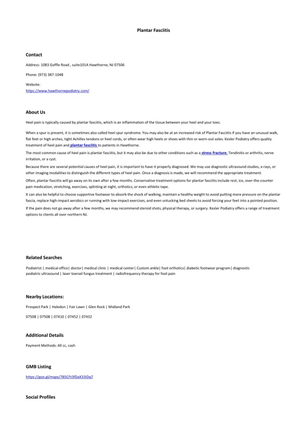
Figure 1 The initial presentation of the foot. There is extensive cyanotic skin changes and bullae formation. Figure 2 Intra-operative photo of the plantar incision and drainage showing extensive myonecrosis with minimal bleeding
Figures 3,4 Appearance of the foot after amputation of the fourth toe prior to the fourth surgical debridement. There is continued necrosis of soft tissue and loss of tissue viability to toes 1-3
Figure 5 After four surgical debridements with amputation of toes 2, 3, and 4, there is now viable bleeding tissue that is free from infection and necrosis Figure 6 The forefoot was disarticulated at the LisFranc joint and the medial and lateral forefoot skin was used as a fasciocutanous flap to cover the defect. http://faoj.org/2008/04/01/necrotizing-soft-tissue-infection-of-the-foot-a-case-report/