Download
1 / 19
190 likes | 1.1k Views
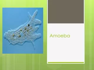
BIO1140 Lab 3: Cellular processes in Amoeba proteus. Objectives of the lab. Identify, photograph and measure organelles found within Amoeba proteus . Observe cell processes such as : amoeboid movement , contractile vacuole cycle and endocytosis .

E N D
Objectives of the lab • Identify, photograph and measure organelles found within Amoeba proteus. • Observe cell processes such as: amoeboid movement, contractile vacuole cycle and endocytosis. • Measure the diameter of the contractile vacuole at selected intervals throughout its cycle. • Use these measurements to plot a graph showing the variation of the vacuole volume during time.
Method (setup) • Carefully connect compound microscope camera to your computer . • Clean optical surfaces of microscope (oculars, objectives, top of condenser and top of lamp housing) using kimwipes.
Microscopic observations • Amoebas are sensitive to light and heat: • Keep the light intensity low during observations.
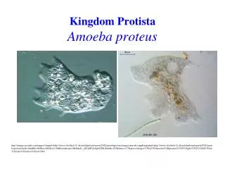
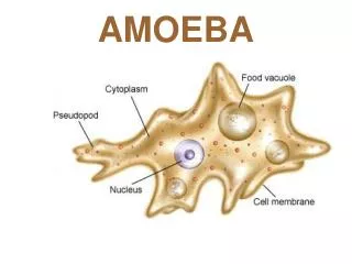
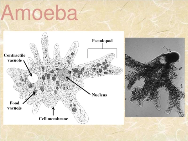
Part I: Amoeba anatomy Identify and know the function of structures and organelles listed in the file: ‘Amoeba structures’ (Lab web site). • Locate an amoeba on your slide using the 4x objective. Then, switch to the 10x and 40x objectives. • There will be questions about Amoeba anatomy and movement in the final exam.
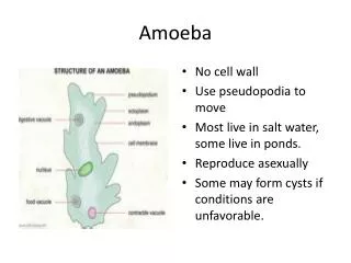
granular endoplasm plasmalemma hyalin cap pseudopodium food vacuole uroid ectoplasm contractile vacuole nucleus dinner
Part II: Amoeboid movement • Your task: Under the 10X observe the formation and elongation of pseudopodia • Switch to the 40x objective to observe the movement of granular endoplasm. • See animation on Digizoo Site • Take several pictures during amoeboid movement • Advice: Take notes, draw sketches and label all structures you observed. Represent the stream of fluid endoplasm using arrows.
Part II: Amoeboid movement TIPS: • Locate an amoeba on the slide using the 4X objective • Adjust the aperture diaphragm (closed = darker with more contrast / open = brighter with less contrast).
Part III: Contractile vacuole cycle Your task: measure the contractile vacuole (CV) diameter throughout its cycle. Contractile vacuole: • Function: osmoregulation and waste removal • Location in the cell: variable • Duration: about 5 minutes at 20°C (cycle duration increases with temperature). • Cycle components: • Diastole (coalescence and continual growth) • Systole (release of CV contents to exterior).
Part III: Contractile vacuole cycle t=0 Systole t=5-8 minutes (300-480s*) t=30 s No vacuole visible yet (diameter=0) Diastole t=60-120 s Small vacuoles start to aggregate and fuse *stop measurements after 480s
Contractile Vacuole: Methods • Locate the contractile vacuole (CV) using the 10x objective. • Once you’ve found a CV switch to 40x objective • Observe at least one full cycle before starting the measurements. • Time is 0 when systole occurs (=vacuole disappears).
Contractile Vacuole: Methods • Take picture of CV every 30 seconds during 2 full cycles (for up to 480 seconds each). • Save your pictures in: K:/BIO1140/BIO1140XX as JPEG files • Warning: SAVE YOU PICTURES PROGRESSIVELY or you’ll lose them. • Once done, measure the diameter of the contractile vacuole (in µm) on each pictures. while measuring, do NOT trace lines on computer screen (-10% immediately)
Contractile Vacuole: Methods • Enter your data into the excel spreadsheet(s) opened on the designated computers • Diameters will be converted into volumes, and class average and standard error will be calculated automatically on the spreadsheet. • Data will be available on the Lab3 page of the lab website (NOTBblearn) tomorrow.
Part IV: Endocytosis • Unmount the Amoeba slide - DO NOT THROW AWAY THE SLIDE -(tech. skills!) • Dispose of coverslips in one of the 2 beakers located a the back of the lab • Go to the endocytosis station • Get a few Amoebas from the flask and transfer them into watch glass • Add 1-2 drops of inducing agent (1% BSA + alcian blue) • Wait 1 minute • Transfer amoebas to the “amoeba slide” with very little liquid (Ask TA) • Add coverslip • Observe shape changes and formation of endocytic canals under microscope
Time Budget • Lab3 Quiz – 10 minutes • TA Prelab Talk - 15 min. • Amoeboid Observation – locomotion & anatomy – 45 minutes • Amoeba contractile vacuole cycle - 75 min. • Endocytosis – 10 min. • Questions to TAs and cleaning of bench 10 min
Lab 3 Report: Title page + Graph Contractile vacuole Graph • Plot CV volume (average +/- standard error) over time (0 to 480 seconds = one cycle) using the class data. • Graph (dot plot) must be done BY HAND (no computer generated graph) on millimeter paper. • Include a caption located below the graph (see lab manual appendix) • Read instruction file on the Lab3 page of lab web • Due date: Week 1 sections: two weeks after the lab Week 2 sections: 1 week after the lab. • Data will be available on lab web sitetomorrow.
Reminders • Clean up or lose technical skill marks • Rinse Amoeba slide and put it back on the tray where you took it. • Dispose of cover slips in the broken glass beaker. • Delete all files your saved • clean up work bench • clean optical surfaces of your microscope • proper microscope return verified by TA before you leave • NEXT LAB: MITOSIS withprelab quiz about lab4
Available documents • Read lab3 documents on the lab website: • List of Amoeba structures • Anatomy scheme • Instruction file for report